

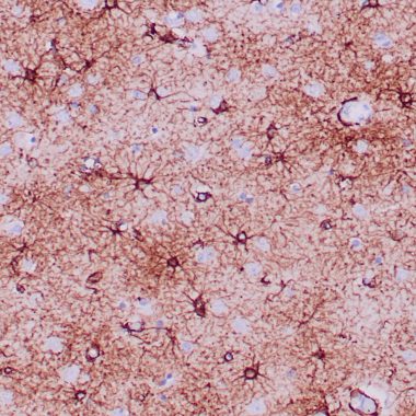
Anti-GFAP antibody (150-250aa) [ZR356] (STJ180514)
SPECIFICATIONS
ClonalityMonoclonal
HostRabbit
ConjugationUnconjugated
IsotypeIgG
ImmunogenRecombinant fragment (around aa 150-250) of human GFAP protein
General Information
| Short Description | Rabbit monoclonal GFAP (150-250aa) antibody for use in IHC-P in human samples. Datasheet included with dilution recommendations, and related reagents. |
| Applications | IHC-P |
| Host | Rabbit |
| Reactivity | Human |
| Note | STRICTLY FOR FURTHER SCIENTIFIC RESEARCH USE ONLY (RUO). MUST NOT TO BE USED IN DIAGNOSTIC OR THERAPEUTIC APPLICATIONS. |
Product Properties
| Clonality | Monoclonal |
| Clone ID | ZR356 |
| Isotype | IgG |
| Conjugation | Unconjugated |
| Purification | Affinity purified |
| Dilution Range | 1:100-200 |
| Formulation | Tris-HCI buffer containing stabilizing protein (BSA) and <0.1% ProClin |
| Storage Instruction | Store at 2‐8°C for up to 24 months. Predilute: Ready to use, no reconstitution necessary. Concentrate: Use dilution range and appropriate lab‐standardized diluent. Stability after dilution: 7 days at 24°C, 3 months at 2‐8°C, 6months at ‐20°C. |
Target Information
| Gene Symbol | GFAP |
| Gene ID | 2670 |
| Uniprot ID | GFAP_HUMAN |
| Immunogen | Recombinant fragment (around aa 150-250) of human GFAP protein |
| Immunogen Region | 150-250aa |
| Specificity | Positive control: Brain or astrocytoma |
Additional Info
| Tissue Specificity | Expressed in cells lacking fibronectin. |
| Post Translational Modifications | Phosphorylated by PKN1. |
| Function | GFAP, a class-III intermediate filament, is a cell-specific marker that, during the development of the central nervous system, distinguishes astrocytes from other glial cells. |
| Protein Name | Glial Fibrillary Acidic ProteinGfap |
| Database Links | Reactome: R-HSA-1251985Reactome: R-HSA-9613829 |
| Cellular Localisation | CytoplasmAssociated With Intermediate Filaments |
| Alternative Antibody Names | Anti-Glial Fibrillary Acidic Protein antibodyAnti-Gfap antibodyAnti-GFAP antibody |
Information sourced from Uniprot.org