• Immunohistochemical analysis of paraffin-embedded Rat-liver tissue. 1, PERK Polyclonal Antibody was diluted at 1:200 (4°C, overnight). 2, Sodium citrate pH 6.0 was used for antibody retrieval (>98°C, 20min). 3, Secondary antibody was diluted at 1:200 (room tempeRature, 30min). Negative control was used by secondary antibody only.
  • Immunohistochemical analysis of paraffin-embedded Rat-lung tissue. 1, PERK Polyclonal Antibody was diluted at 1:200 (4°C, overnight). 2, Sodium citrate pH 6.0 was used for antibody retrieval (>98°C, 20min). 3, Secondary antibody was diluted at 1:200 (room tempeRature, 30min). Negative control was used by secondary antibody only.
  • Immunohistochemical analysis of paraffin-embedded Human-Appendix tissue. 1, PERK Polyclonal Antibody was diluted at 1:200 (4°C, overnight). 2, Sodium citrate pH 6.0 was used for antibody retrieval (>98°C, 20min). 3, Secondary antibody was diluted at 1:200 (room tempeRature, 30min). Negative control was used by secondary antibody only.
  • Immunohistochemical analysis of paraffin-embedded Rat-heart tissue. 1, PERK Polyclonal Antibody was diluted at 1:200 (4°C, overnight). 2, Sodium citrate pH 6.0 was used for antibody retrieval (>98°C, 20min). 3, Secondary antibody was diluted at 1:200 (room tempeRature, 30min). Negative control was used by secondary antibody only.
  • Immunohistochemistry analysis of paraffin-embedded human colon carcinoma, using PEK/PERK Antibody. The picture on the right is blocked with the synthesized peptide.
  • Immunofluorescence analysis of rat-lung tissue. 1, PERK Polyclonal Antibody (red) was diluted at 1:200 (4°C, overnight). 2, Cy3 labled Secondary antibody was diluted at 1:300 (room temperature, 50min).3, Picture B: DAPI (blue) 10min. Picture A:Target. Picture B: DAPI. Picture C: merge of A+B
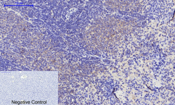
  • Immunohistochemical analysis of paraffin-embedded Human-Tonsil tissue. 1, PERK Polyclonal Antibody was diluted at 1:200 (4°C, overnight). 2, Sodium citrate pH 6.0 was used for antibody retrieval (>98°C, 20min). 3, Secondary antibody was diluted at 1:200 (room tempeRature, 30min). Negative control was used by secondary antibody only.
  • Immunohistochemical analysis of paraffin-embedded Mouse-liver tissue. 1, PERK Polyclonal Antibody was diluted at 1:200 (4°C, overnight). 2, Sodium citrate pH 6.0 was used for antibody retrieval (>98°C, 20min). 3, Secondary antibody was diluted at 1:200 (room tempeRature, 30min). Negative control was used by secondary antibody only.
  • Immunofluorescence analysis of mouse-lung tissue. 1, PERK Polyclonal Antibody (red) was diluted at 1:200 (4°C, overnight). 2, Cy3 labled Secondary antibody was diluted at 1:300 (room temperature, 50min).3, Picture B: DAPI (blue) 10min. Picture A:Target. Picture B: DAPI. Picture C: merge of A+B
  • Immunofluorescence analysis of mouse-lung tissue. 1, PERK Polyclonal Antibody (red) was diluted at 1:200 (4°C, overnight). 2, Cy3 labled Secondary antibody was diluted at 1:300 (room temperature, 50min).3, Picture B: DAPI (blue) 10min. Picture A:Target. Picture B: DAPI. Picture C: merge of A+B
  • Immunofluorescence analysis of rat-spleen tissue. 1, PERK Polyclonal Antibody (red) was diluted at 1:200 (4°C, overnight). 2, Cy3 labled Secondary antibody was diluted at 1:300 (room temperature, 50min).3, Picture B: DAPI (blue) 10min. Picture A:Target. Picture B: DAPI. Picture C: merge of A+B
  • Immunofluorescence analysis of rat-spleen tissue. 1, PERK Polyclonal Antibody (red) was diluted at 1:200 (4°C, overnight). 2, Cy3 labled Secondary antibody was diluted at 1:300 (room temperature, 50min).3, Picture B: DAPI (blue) 10min. Picture A:Target. Picture B: DAPI. Picture C: merge of A+B
  • Immunofluorescence analysis of rat-lung tissue. 1, PERK Polyclonal Antibody (red) was diluted at 1:200 (4°C, overnight). 2, Cy3 labled Secondary antibody was diluted at 1:300 (room temperature, 50min).3, Picture B: DAPI (blue) 10min. Picture A:Target. Picture B: DAPI. Picture C: merge of A+B
  • Immunohistochemical analysis of paraffin-embedded Rat-kidney tissue. 1, PERK Polyclonal Antibody was diluted at 1:200 (4°C, overnight). 2, Sodium citrate pH 6.0 was used for antibody retrieval (>98°C, 20min). 3, Secondary antibody was diluted at 1:200 (room tempeRature, 30min). Negative control was used by secondary antibody only.
  • Immunohistochemical analysis of paraffin-embedded Human-lung tissue. 1, PERK Polyclonal Antibody was diluted at 1:200 (4°C, overnight). 2, Sodium citrate pH 6.0 was used for antibody retrieval (>98°C, 20min). 3, Secondary antibody was diluted at 1:200 (room tempeRature, 30min). Negative control was used by secondary antibody only.
  • Immunohistochemical analysis of paraffin-embedded Mouse-heart tissue. 1, PERK Polyclonal Antibody was diluted at 1:200 (4°C, overnight). 2, Sodium citrate pH 6.0 was used for antibody retrieval (>98°C, 20min). 3, Secondary antibody was diluted at 1:200 (room tempeRature, 30min). Negative control was used by secondary antibody only.
  • Immunohistochemical analysis of paraffin-embedded Human-stomach tissue. 1, PERK Polyclonal Antibody was diluted at 1:200 (4°C, overnight). 2, Sodium citrate pH 6.0 was used for antibody retrieval (>98°C, 20min). 3, Secondary antibody was diluted at 1:200 (room tempeRature, 30min). Negative control was used by secondary antibody only.
  • Immunohistochemical analysis of paraffin-embedded Mouse-lung tissue. 1, PERK Polyclonal Antibody was diluted at 1:200 (4°C, overnight). 2, Sodium citrate pH 6.0 was used for antibody retrieval (>98°C, 20min). 3, Secondary antibody was diluted at 1:200 (room tempeRature, 30min). Negative control was used by secondary antibody only.
  • Immunohistochemical analysis of paraffin-embedded Human-stomach-cancer tissue. 1, PERK Polyclonal Antibody was diluted at 1:200 (4°C, overnight). 2, Sodium citrate pH 6.0 was used for antibody retrieval (>98°C, 20min). 3, Secondary antibody was diluted at 1:200 (room tempeRature, 30min). Negative control was used by secondary antibody only.
  • Western blot analysis of lysates from MCF-7 cells, using PEK/PERK Antibody. The lane on the right is blocked with the synthesized peptide.
  • Immunofluorescence analysis of Hela cell. 1, PERK Polyclonal Antibody (green) was diluted at 1:200 (4°C overnight). 2, Goat Anti Rabbit Alexa Fluor 488 Catalog: (NA was diluted at 1:1000 (room temperature, 50min). 3 DAPI (blue) 10min.

Anti-EIF2AK3 antibody (947-996 aa) (STJ95031)

SKU:
STJ95031

Current Stock:
Host: Rabbit
Applications: IF/WB/IHC/ELISA
Reactivity: Human/Mouse/Rat
Note: STRICTLY FOR FURTHER SCIENTIFIC RESEARCH USE ONLY (RUO). MUST NOT TO BE USED IN DIAGNOSTIC OR THERAPEUTIC APPLICATIONS.
Short Description: Rabbit polyclonal antibody anti-Eukaryotic translation initiation factor 2-alpha kinase 3 (947-996 aa) is suitable for use in Immunofluorescence, Western Blot, Immunohistochemistry and ELISA research applications.
Clonality: Polyclonal
Conjugation: Unconjugated
Isotype: IgG
Formulation: Liquid in PBS containing 50% Glycerol, 0.5% BSA and 0.02% Sodium Azide.
Purification: The antibody was affinity-purified from rabbit antiserum by affinity-chromatography using epitope-specific immunogen.
Concentration: 1 mg/mL
Dilution Range: IF 1:50-200
WB 1:500-1:2000
IHC 1:100-1:300
ELISA 1:40000
Storage Instruction: Store at-20°C for up to 1 year from the date of receipt, and avoid repeat freeze-thaw cycles.
Gene Symbol: EIF2AK3
Gene ID: 9451
Uniprot ID: E2AK3_HUMAN
Immunogen Region: 947-996 aa
Specificity: PERK Polyclonal Antibody detects endogenous levels of PERK protein.
Immunogen: The antiserum was produced against synthesized peptide derived from the human EIF2AK3 at the amino acid range 947-996
Post Translational Modifications Oligomerization of the N-terminal ER luminal domain by ER stress promotes EIF2AK3/PERK trans-autophosphorylation of the C-terminal cytoplasmic kinase domain at multiple residues including Thr-982 on the kinase activation loop. Autophosphorylated at Tyr-619 following endoplasmic reticulum stress, leading to activate its activity. Dephosphorylated at Tyr-619 by PTPN1/PTP1B, leading to inactivate its enzyme activity. Phosphorylation at Thr-802 by AKT (AKT1, AKT2 and/or AKT3) inactivates EIF2AK3/PERK. ADP-ribosylated by PARP16 upon ER stress, which increases kinase activity.
Function Metabolic-stress sensing protein kinase that phosphorylates the alpha subunit of eukaryotic translation initiation factor 2 (EIF2S1/eIF-2-alpha) in response to various stress, such as unfolded protein response (UPR). Key effector of the integrated stress response (ISR) to unfolded proteins: EIF2AK3/PERK specifically recognizes and binds misfolded proteins, leading to its activation and EIF2S1/eIF-2-alpha phosphorylation. EIF2S1/eIF-2-alpha phosphorylation in response to stress converts EIF2S1/eIF-2-alpha in a global protein synthesis inhibitor, leading to a global attenuation of cap-dependent translation, while concomitantly initiating the preferential translation of ISR-specific mRNAs, such as the transcriptional activators ATF4 and QRICH1, and hence allowing ATF4- and QRICH1-mediated reprogramming. The EIF2AK3/PERK-mediated unfolded protein response increases mitochondrial oxidative phosphorylation by promoting ATF4-mediated expression of COX7A2L/SCAF1, thereby increasing formation of respiratory chain supercomplexes. In contrast to most subcellular compartments, mitochondria are protected from the EIF2AK3/PERK-mediated unfolded protein response due to EIF2AK3/PERK inhibition by ATAD3A at mitochondria-endoplasmic reticulum contact sites. In addition to EIF2S1/eIF-2-alpha, also phosphorylates NFE2L2/NRF2 in response to stress, promoting release of NFE2L2/NRF2 from the BCR(KEAP1) complex, leading to nuclear accumulation and activation of NFE2L2/NRF2. Serves as a critical effector of unfolded protein response (UPR)-induced G1 growth arrest due to the loss of cyclin-D1 (CCND1). Involved in control of mitochondrial morphology and function.
Protein Name Eukaryotic Translation Initiation Factor 2-Alpha Kinase 3
Prkr-Like Endoplasmic Reticulum Kinase
Pancreatic Eif2-Alpha Kinase
Hspek
Protein Tyrosine Kinase Eif2ak3
Database Links Reactome: R-HSA-381042
Reactome: R-HSA-9700645
Reactome: R-HSA-9725370
Reactome: R-HSA-9755511
Cellular Localisation Endoplasmic Reticulum Membrane
Single-Pass Type I Membrane Protein
Localizes To The Localizes To Endoplasmic Reticulum Membrane
Also Present At Mitochondria-Endoplasmic Reticulum Contact Sites
Where It Interacts With Atad3a
Alternative Antibody Names Anti-Eukaryotic Translation Initiation Factor 2-Alpha Kinase 3 antibody
Anti-Prkr-Like Endoplasmic Reticulum Kinase antibody
Anti-Pancreatic Eif2-Alpha Kinase antibody
Anti-Hspek antibody
Anti-Protein Tyrosine Kinase Eif2ak3 antibody
Anti-EIF2AK3 antibody
Anti-PEK antibody
Anti-PERK antibody

Information sourced from Uniprot.org

12 months for antibodies. 6 months for ELISA Kits. Please see website T&Cs for further guidance