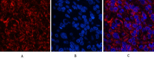
• Immunofluorescence analysis of Human-lung-cancer tissue. 1, Cleaved-Caspase-3 p12 (D175) Polyclonal Antibody (red) was diluted at 1:200 (4°C, overnight). 2, Cy3 labled Secondary antibody was diluted at 1:300 (room temperature, 50min).3, Picture B: DAPI (blue) 10min. Picture A:Target. Picture B: DAPI. Picture C: merge of A+B
  • Western blot analysis of various cells using Cleaved-Caspase-3 p12 (D175) Polyclonal Antibody diluted at 1:1000
  • Immunofluorescence analysis of Human-lung-cancer tissue. 1, Cleaved-Caspase-3 p12 (D175) Polyclonal Antibody (red) was diluted at 1:200 (4°C, overnight). 2, Cy3 labled Secondary antibody was diluted at 1:300 (room temperature, 50min).3, Picture B: DAPI (blue) 10min. Picture A:Target. Picture B: DAPI. Picture C: merge of A+B
  • Immunofluorescence analysis of Human-lung-cancer tissue. 1, Cleaved-Caspase-3 p12 (D175) Polyclonal Antibody (red) was diluted at 1:200 (4°C, overnight). 2, Cy3 labled Secondary antibody was diluted at 1:300 (room temperature, 50min).3, Picture B: DAPI (blue) 10min. Picture A:Target. Picture B: DAPI. Picture C: merge of A+B
  • Immunohistochemical analysis of paraffin-embedded Human-breast-cancer tissue. 1, Cleaved-Caspase-3 p12 (D175) Polyclonal Antibody was diluted at 1:200 (4°C, overnight). 2, Sodium citrate pH 6.0 was used for antibody retrieval (>98°C, 20min). 3, Secondary antibody was diluted at 1:200 (room tempeRature, 30min). Negative control was used by secondary antibody only.
  • Immunofluorescence analysis of Human-liver-cancer tissue. 1, Cleaved-Caspase-3 p12 (D175) Polyclonal Antibody (red) was diluted at 1:200 (4°C, overnight). 2, Cy3 labled Secondary antibody was diluted at 1:300 (room temperature, 50min).3, Picture B: DAPI (blue) 10min. Picture A:Target. Picture B: DAPI. Picture C: merge of A+B
  • Immunofluorescence analysis of Human-kidney-cancer tissue. 1, Cleaved-Caspase-3 p12 (D175) Polyclonal Antibody (red) was diluted at 1:200 (4°C, overnight). 2, Cy3 labled Secondary antibody was diluted at 1:300 (room temperature, 50min).3, Picture B: DAPI (blue) 10min. Picture A:Target. Picture B: DAPI. Picture C: merge of A+B
  • Immunohistochemistry analysis of paraffin-embedded human brain tissue, using Caspase 3 (Cleaved-Asp175) Antibody. The picture on the right is blocked with the synthesized peptide.
  • Immunohistochemical analysis of paraffin-embedded Human-kidney tissue. 1, Cleaved-Caspase-3 p12 (D175) Polyclonal Antibody was diluted at 1:200 (4°C, overnight). 2, Sodium citrate pH 6.0 was used for antibody retrieval (>98°C, 20min). 3, Secondary antibody was diluted at 1:200 (room tempeRature, 30min). Negative control was used by secondary antibody only.
  • Immunohistochemical analysis of paraffin-embedded Human-stomach-cancer tissue. 1, Cleaved-Caspase-3 p12 (D175) Polyclonal Antibody was diluted at 1:200 (4°C, overnight). 2, Sodium citrate pH 6.0 was used for antibody retrieval (>98°C, 20min). 3, Secondary antibody was diluted at 1:200 (room tempeRature, 30min). Negative control was used by secondary antibody only.
  • Immunohistochemical analysis of paraffin-embedded Human-lung-cancer tissue. 1, Cleaved-Caspase-3 p12 (D175) Polyclonal Antibody was diluted at 1:200 (4°C, overnight). 2, Sodium citrate pH 6.0 was used for antibody retrieval (>98°C, 20min). 3, Secondary antibody was diluted at 1:200 (room tempeRature, 30min). Negative control was used by secondary antibody only.
  • Immunohistochemical analysis of paraffin-embedded Human-liver-cancer tissue. 1, Cleaved-Caspase-3 p12 (D175) Polyclonal Antibody was diluted at 1:200 (4°C, overnight). 2, Sodium citrate pH 6.0 was used for antibody retrieval (>98°C, 20min). 3, Secondary antibody was diluted at 1:200 (room tempeRature, 30min). Negative control was used by secondary antibody only.
  • Immunohistochemical analysis of paraffin-embedded Human-liver tissue. 1, Cleaved-Caspase-3 p12 (D175) Polyclonal Antibody was diluted at 1:200 (4°C, overnight). 2, Sodium citrate pH 6.0 was used for antibody retrieval (>98°C, 20min). 3, Secondary antibody was diluted at 1:200 (room tempeRature, 30min). Negative control was used by secondary antibody only.
  • Western blot analysis of KB cells using Cleaved-Caspase-3 p12 (D175) Polyclonal Antibody diluted at 1:1000
  • Immunofluorescence analysis of Human-liver-cancer tissue. 1, Cleaved-Caspase-3 p12 (D175) Polyclonal Antibody (red) was diluted at 1:200 (4°C, overnight). 2, Cy3 labled Secondary antibody was diluted at 1:300 (room temperature, 50min).3, Picture B: DAPI (blue) 10min. Picture A:Target. Picture B: DAPI. Picture C: merge of A+B
  • Immunohistochemical analysis of paraffin-embedded Human lung cancer. Antibody was diluted at 1:100 (4°C overnight). High-pressure and temperature Tris-EDTA, pH8.0 was used for antigen retrieval. Negetive contrl (right) obtaned from antibody was pre-absorbed by immunogen peptide.
  • Immunofluorescence analysis of Human-liver-cancer tissue. 1, Cleaved-Caspase-3 p12 (D175) Polyclonal Antibody (red) was diluted at 1:200 (4°C, overnight). 2, Cy3 labled Secondary antibody was diluted at 1:300 (room temperature, 50min).3, Picture B: DAPI (blue) 10min. Picture A:Target. Picture B: DAPI. Picture C: merge of A+B
  • Western blot analysis of lysates from HeLa cells, treated with Etoposide 25uM 60', using Caspase 3 (Cleaved-Asp175) Antibody. The lane on the right is blocked with the synthesized peptide.
  • Immunohistochemical analysis of paraffin-embedded Human-breast tissue. 1, Cleaved-Caspase-3 p12 (D175) Polyclonal Antibody was diluted at 1:200 (4°C, overnight). 2, Sodium citrate pH 6.0 was used for antibody retrieval (>98°C, 20min). 3, Secondary antibody was diluted at 1:200 (room tempeRature, 30min). Negative control was used by secondary antibody only.

Anti-Cleaved-CASP3-D175 antibody (157-206 aa) (STJ90003)

SKU:
STJ90003

Current Stock:
Host: Rabbit
Applications: WB/IF/IHC/ELISA
Reactivity: Human/Rat/Mouse
Note: STRICTLY FOR FURTHER SCIENTIFIC RESEARCH USE ONLY (RUO). MUST NOT TO BE USED IN DIAGNOSTIC OR THERAPEUTIC APPLICATIONS.
Short Description: Rabbit polyclonal antibody anti-Cleaved-Caspase-3-D175 (157-206 aa) is suitable for use in Western Blot, Immunofluorescence, Immunohistochemistry and ELISA research applications.
Clonality: Polyclonal
Conjugation: Unconjugated
Isotype: IgG
Formulation: Liquid in PBS containing 50% Glycerol, 0.5% BSA and 0.02% Sodium Azide.
Purification: The antibody was affinity-purified from rabbit antiserum by affinity-chromatography using epitope-specific immunogen.
Concentration: 1 mg/mL
Dilution Range: WB 1:500-2000
IHC-P 1:50-300
IF 1:50-300
Storage Instruction: Store at-20°C for up to 1 year from the date of receipt, and avoid repeat freeze-thaw cycles.
Gene Symbol: CASP3
Gene ID: 836
Uniprot ID: CASP3_HUMAN
Immunogen Region: 157-206 aa
Specificity: Cleaved-Caspase-3 p12 (D175) Polyclonal Antibody detects endogenous levels of fragment of activated Caspase-3 p12 protein resulting from cleavage adjacent to D175.
Immunogen: The antiserum was produced against synthesized peptide derived from the human Caspase 3 at the amino acid range 157-206
Post Translational Modifications Cleavage by granzyme B, caspase-6, caspase-8 and caspase-10 generates the two active subunits. Additional processing of the propeptides is likely due to the autocatalytic activity of the activated protease. Active heterodimers between the small subunit of caspase-7 protease and the large subunit of caspase-3 also occur and vice versa. S-nitrosylated on its catalytic site cysteine in unstimulated human cell lines and denitrosylated upon activation of the Fas apoptotic pathway, associated with an increase in intracellular caspase activity. Fas therefore activates caspase-3 not only by inducing the cleavage of the caspase zymogen to its active subunits, but also by stimulating the denitrosylation of its active site thiol. (Microbial infection) ADP-riboxanation by C.violaceum CopC blocks CASP3 processing, preventing CASP3 activation and ability to recognize and cleave substrates.
Function Thiol protease that acts as a major effector caspase involved in the execution phase of apoptosis. Following cleavage and activation by initiator caspases (CASP8, CASP9 and/or CASP10), mediates execution of apoptosis by catalyzing cleavage of many proteins. At the onset of apoptosis, it proteolytically cleaves poly(ADP-ribose) polymerase PARP1 at a '216-Asp-|-Gly-217' bond. Cleaves and activates sterol regulatory element binding proteins (SREBPs) between the basic helix-loop-helix leucine zipper domain and the membrane attachment domain. Cleaves and activates caspase-6, -7 and -9. Involved in the cleavage of huntingtin. Triggers cell adhesion in sympathetic neurons through RET cleavage. Cleaves and inhibits serine/threonine-protein kinase AKT1 in response to oxidative stress. Acts as an inhibitor of type I interferon production during virus-induced apoptosis by mediating cleavage of antiviral proteins CGAS, IRF3 and MAVS, thereby preventing cytokine overproduction. Also involved in pyroptosis by mediating cleavage and activation of gasdermin-E (GSDME). Cleaves XRCC4 and phospholipid scramblase proteins XKR4, XKR8 and XKR9, leading to promote phosphatidylserine exposure on apoptotic cell surface.
Protein Name Caspase-3
Casp-3
Apopain
Cysteine Protease Cpp32
Cpp-32
Protein Yama
Srebp Cleavage Activity 1
Sca-1 Cleaved Into - Caspase-3 Subunit P17 - Caspase-3 Subunit P12
Database Links Reactome: R-HSA-111459
Reactome: R-HSA-111463
Reactome: R-HSA-111464
Reactome: R-HSA-111465
Reactome: R-HSA-111469
Reactome: R-HSA-140342
Reactome: R-HSA-1474228
Reactome: R-HSA-2028269
Reactome: R-HSA-205025
Reactome: R-HSA-211736
Reactome: R-HSA-264870
Reactome: R-HSA-351906
Reactome: R-HSA-418889
Reactome: R-HSA-449836
Reactome: R-HSA-5620971
Cellular Localisation Cytoplasm
Alternative Antibody Names Anti-Caspase-3 antibody
Anti-Casp-3 antibody
Anti-Apopain antibody
Anti-Cysteine Protease Cpp32 antibody
Anti-Cpp-32 antibody
Anti-Protein Yama antibody
Anti-Srebp Cleavage Activity 1 antibody
Anti-Sca-1 Cleaved Into - Caspase-3 Subunit P17 - Caspase-3 Subunit P12 antibody
Anti-CASP3 antibody
Anti-CPP32 antibody

Information sourced from Uniprot.org

12 months for antibodies. 6 months for ELISA Kits. Please see website T&Cs for further guidance