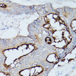
| Tissue Specificity | Expressed in columnar epithelial cells of the colon (at protein level). The predominant forms expressed by T cells are those containing a long cytoplasmic domain. Expressed in granulocytes and lymphocytes. Leukocytes only express isoforms 6 and isoform 1. |
| Post Translational Modifications | Isoform 1: Phosphorylated on serine and tyrosine. Isoform 1 is phosphorylated on tyrosine by Src family kinases like SRC and LCK and by receptor like CSF3R, EGFR and INSR upon stimulation. Phosphorylated at Ser-508.mediates activity. Phosphorylated at Tyr-493.regulates activity. Phosphorylated at Tyr-493 by EGFR and INSR upon stimulation.this phosphorylation is Ser-508-phosphorylation-dependent.mediates cellular internalization.increases interaction with downstream proteins like SHC1 and FASN. Phosphorylated at Tyr-493 and Tyr-520 by LCK.mediates PTPN6 association and is regulated by homophilic ligation of CEACAM1 in the absence of T cell activation. Phosphorylated at Tyr-520.mediates interaction with PTPN11. Isoform 8: Phosphorylated on serine and threonine. |
| Function | Isoform 1: Cell adhesion protein that mediates homophilic cell adhesion in a calcium-independent manner. Plays a role as coinhibitory receptor in immune response, insulin action and also functions as an activator during angiogenesis. Its coinhibitory receptor function is phosphorylation- and PTPN6 -dependent, which in turn, suppress signal transduction of associated receptors by dephosphorylation of their downstream effectors. Plays a role in immune response, of T cells, natural killer (NK) and neutrophils. Upon TCR/CD3 complex stimulation, inhibits TCR-mediated cytotoxicity by blocking granule exocytosis by mediating homophilic binding to adjacent cells, allowing interaction with and phosphorylation by LCK and interaction with the TCR/CD3 complex which recruits PTPN6 resulting in dephosphorylation of CD247 and ZAP70. Also inhibits T cell proliferation and cytokine production through inhibition of JNK cascade and plays a crucial role in regulating autoimmunity and anti-tumor immunity by inhibiting T cell through its interaction with HAVCR2. Upon natural killer (NK) cells activation, inhibit KLRK1-mediated cytolysis of CEACAM1-bearing tumor cells by trans-homophilic interactions with CEACAM1 on the target cell and lead to cis-interaction between CEACAM1 and KLRK1, allowing PTPN6 recruitment and then VAV1 dephosphorylation. Upon neutrophils activation negatively regulates IL1B production by recruiting PTPN6 to a SYK-TLR4-CEACAM1 complex, that dephosphorylates SYK, reducing the production of reactive oxygen species (ROS) and lysosome disruption, which in turn, reduces the activity of the inflammasome. Down-regulates neutrophil production by acting as a coinhibitory receptor for CSF3R by down-regulating the CSF3R-STAT3 pathway through recruitment of PTPN6 that dephosphorylates CSF3R. Also regulates insulin action by promoting INS clearance and regulating lipogenesis in liver through regulating insulin signaling. Upon INS stimulation, undergoes phosphorylation by INSR leading to INS clearance by increasing receptor-mediated insulin endocytosis. This inernalization promotes interaction with FASN leading to receptor-mediated insulin degradation and to reduction of FASN activity leading to negative regulation of fatty acid synthesis. INSR-mediated phosphorylation also provokes a down-regulation of cell proliferation through SHC1 interaction resulting in decrease coupling of SHC1 to the MAPK3/ERK1-MAPK1/ERK2 and phosphatidylinositol 3-kinase pathways. Functions as activator in angiogenesis by promoting blood vessel remodeling through endothelial cell differentiation and migration and in arteriogenesis by increasing the number of collateral arteries and collateral vessel calibers after ischemia. Also regulates vascular permeability through the VEGFR2 signaling pathway resulting in control of nitric oxide production. Down-regulates cell growth in response to EGF through its interaction with SHC1 that mediates interaction with EGFR resulting in decrease coupling of SHC1 to the MAPK3/ERK1-MAPK1/ERK2 pathway. Negatively regulates platelet aggregation by decreasing platelet adhesion on type I collagen through the GPVI-FcRgamma complex. Inhibits cell migration and cell scattering through interaction with FLNA.interferes with the interaction of FLNA with RALA. Mediates bile acid transport activity in a phosphorylation dependent manner. Negatively regulates osteoclastogenesis. Isoform 8: Cell adhesion protein that mediates homophilic cell adhesion in a calcium-independent manner. Promotes populations of T cells regulating IgA production and secretion associated with control of the commensal microbiota and resistance to enteropathogens. |
| Protein Name | Cell Adhesion Molecule Ceacam1Biliary Glycoprotein 1Bgp-1Carcinoembryonic Antigen-Related Cell Adhesion Molecule 1Cea Cell Adhesion Molecule 1Cd Antigen Cd66a |
| Database Links | Reactome: R-HSA-1566977Reactome: R-HSA-202733Reactome: R-HSA-6798695Reactome: R-HSA-9854909 |
| Cellular Localisation | Isoform 1: Cell MembraneSingle-Pass Type I Membrane ProteinLateral Cell MembraneApical Cell MembraneBasal Cell MembraneCell JunctionAdherens JunctionCanalicular Domain Of Hepatocyte Plasma MembranesFound As A Mixture Of MonomerDimer And Oligomer In The Plasma MembraneOccurs Predominantly As Cis-Dimers And/Or Small Cis-Oligomers In The Cell Junction RegionsFound As Dimer In The SolutionPredominantly Localized To The Lateral Cell MembranesIsoform 2: SecretedIsoform 3: SecretedIsoform 4: SecretedIsoform 5: Cell MembraneIsoform 6: Cell MembraneIsoform 7: Cell MembraneIsoform 8: Cell MembraneCytoplasmic VesicleSecretory Vesicle MembraneCo-Localizes With Anxa2 In Secretory Vesicles And With S100a10/P11 At The Plasma MembraneCell ProjectionMicrovillus MembraneLocalized To The Apical Glycocalyx SurfaceColocalizes With Ceacam20 At The Apical Brush Border Of Intestinal Cells |
| Alternative Antibody Names | Anti-Cell Adhesion Molecule Ceacam1 antibodyAnti-Biliary Glycoprotein 1 antibodyAnti-Bgp-1 antibodyAnti-Carcinoembryonic Antigen-Related Cell Adhesion Molecule 1 antibodyAnti-Cea Cell Adhesion Molecule 1 antibodyAnti-Cd Antigen Cd66a antibodyAnti-CEACAM1 antibodyAnti-BGP antibodyAnti-BGP1 antibody |
Information sourced from Uniprot.org


![Anti-CEACAM1 antibody [SM5076] (STJA0005076) Anti-CEACAM1 antibody [SM5076] (STJA0005076)](https://cdn11.bigcommerce.com/s-zso2xnchw9/images/stencil/600x533/products/180131/403420/STJA0005076_1__02499.1738950476.png?c=1)
![Anti-CEACAM1 antibody [SM4958] (STJA0004958) Anti-CEACAM1 antibody [SM4958] (STJA0004958)](https://cdn11.bigcommerce.com/s-zso2xnchw9/images/stencil/600x533/products/180029/403272/STJA0004958_1__54975.1738950350.png?c=1)
