


Anti-CD63 antibody (120aa to 175aa) (STJ140029)
SPECIFICATIONS
ClonalityPolyclonal
HostGoat
ConjugationUnconjugated
IsotypeIgG
ImmunogenPurified recombinant peptide derived from within residues 120 aa to 175 aa of human CD63 produced in E. coli.
General Information
| Short Description | Goat polyclonal Lysosome-associated membrane glycoprotein 3 (120aa to 175aa) antibody for use in WB, IF, IHC-P and IHC-F in human, mouse, rat, bovine, canine, chicken, avian, donkey, feline, goat, guinea pig, hamster, horse, porcine, rabbit, sheep an |
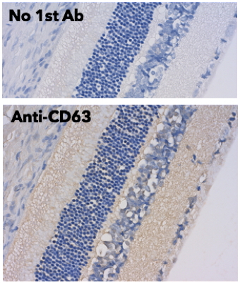
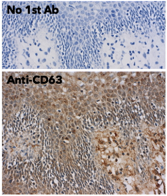
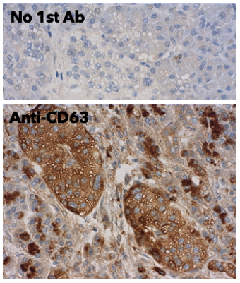
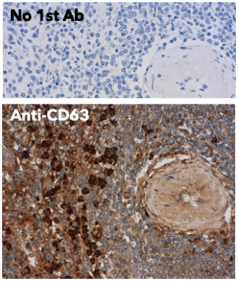
| Applications | WB/IF/IHC-P/IHC-F |
| Host | Goat |
| Reactivity | Human/Mouse/Rat/Bovine/Canine/Chicken/Avian/Donkey/Feline/Goat/Guinea Pig/Hamster/Horse/Porcine/Rabbit/Sheep/Simian |
| Note | STRICTLY FOR FURTHER SCIENTIFIC RESEARCH USE ONLY (RUO). MUST NOT TO BE USED IN DIAGNOSTIC OR THERAPEUTIC APPLICATIONS. |
Product Properties
| Clonality | Polyclonal |
| Isotype | IgG |
| Conjugation | Unconjugated |
| Concentration | 3 mg/mL |
| Purification | This antibody is epitope-affinity purified from goat antiserum. |
| Dilution Range | WB 1:500-1:5000IF 1:25-1:250IHC-F 1:250-1:1000IHC-P 1:250-1:1000Amorim M Martins B Caramelo F et al. Front Med-Lausanne 2022 May PMID: 35692536Matsui T Sakamaki Y Nakashima S et al. Cell Rep 2022 May PMID: 35649370Martin |
| Formulation | PBS, 20% Glycerol and 0.05% Sodium Azide. |
| Storage Instruction | For continuous use, store at 2-8 C for one-two days. For extended storage, store in-20 C freezer. Working dilution samples should be discarded if not used within 12 hours. |
Target Information
| Gene Symbol | LAMP3 |
| Gene ID | 27074 |
| Uniprot ID | LAMP3_HUMAN |
| Accession Number | ENSG00000135404 |
| Immunogen | Purified recombinant peptide derived from within residues 120 aa to 175 aa of human CD63 produced in E. coli. |
| Immunogen Region | 120aa to 175aa |
| Immunogen Sequence | MENYPKNNHTASILDRMQAD ENYPKNNHTASILDRMQADF NYPKNNHTASILDRMQADFK |
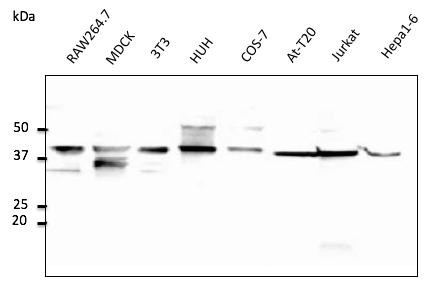
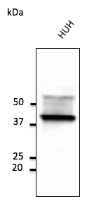
| Specificity | Reacts with CD63, a 40-60 kDa glycoprotein, detected by Western blot in the following human (HeLa, HUH, Jurkat) , mouse (AtT-20, Hepa, 3T3, RAW264.7) , canine (MDCK) and monkey (COS-7) whole cell lysates. |
Additional Info
| Tissue Specificity | Detected in tonsil interdigitating dendritic cells, in spleen, lymph node, Peyer's patches in the small instestine, in thymus medulla and in B-cells (at protein level). Expressed in lymphoid organs and dendritic cells. Expressed in lung. Up-regulated in carcinomas of the esophagus, colon, rectum, ureter, stomach, breast, fallopian tube, thyroid and parotid tissues. |
| Function | Lysosomal membrane glycoprotein which plays a role in the unfolded protein response (UPR) that contributes to protein degradation and cell survival during proteasomal dysfunction. Plays a role in the process of fusion of the lysosome with the autophagosome, thereby modulating the autophagic process. Promotes hepatocellular lipogenesis through activation of the PI3K/Akt pathway. May also play a role in dendritic cell function and in adaptive immunity. (Microbial infection) Plays a positive role in post-entry steps of influenza A virus replication, either virus uncoating, cytosolic transport, or nuclear import of viral components, and promotes nuclear accumulation of influenza nucleoprotein/NP at early stages of viral infection. (Microbial infection) Supports the FURIN-mediated cleavage of mumps virus fusion protein F by interacting with both FURIN and the unprocessed form but not the processed form of the viral protein F. (Microbial infection) Promotes the intracellular proliferation of Salmonella typhimuium. |
| Protein Name | Lysosome-Associated Membrane Glycoprotein 3Lamp-3Lysosomal-Associated Membrane Protein 3Dc-Lysosome-Associated Membrane GlycoproteinDc LampProtein Tsc403Cd Antigen Cd208 |
| Database Links | |
| Cellular Localisation | Cell SurfaceLysosome MembraneSingle-Pass Type I Membrane ProteinCytoplasmic Vesicle MembraneEarly Endosome MembraneDuring Dendritic Cell MaturationDetected On Cytoplasmic Vesicles (The Mhc Ii Compartment) That Contain Mhc Ii ProteinsLamp1Lamp2 And Lamp3Detected On Lysosomes In Mature Dendritic Cells |
| Alternative Antibody Names | Anti-Lysosome-Associated Membrane Glycoprotein 3 antibodyAnti-Lamp-3 antibodyAnti-Lysosomal-Associated Membrane Protein 3 antibodyAnti-Dc-Lysosome-Associated Membrane Glycoprotein antibodyAnti-Dc Lamp antibodyAnti-Protein Tsc403 antibodyAnti-Cd Antigen Cd208 antibodyAnti-LAMP3 antibodyAnti-DCLAMP antibodyAnti-TSC403 antibody |
Information sourced from Uniprot.org