

Anti-Caldesmon antibody (Full length) [ZM79] (STJ180318)
SPECIFICATIONS
ClonalityMonoclonal
HostMouse
ConjugationUnconjugated
IsotypeIgG1k
ImmunogenRecombinant full-length human CALD1 protein
General Information
| Short Description | Mouse monoclonal Caldesmon (Full length) antibody for use in IHC-P in human samples. Datasheet included with dilution recommendations, and related reagents. |
| Applications | IHC-P |
| Host | Mouse |
| Reactivity | Human |
| Note | STRICTLY FOR FURTHER SCIENTIFIC RESEARCH USE ONLY (RUO). MUST NOT TO BE USED IN DIAGNOSTIC OR THERAPEUTIC APPLICATIONS. |
Product Properties
| Clonality | Monoclonal |
| Clone ID | ZM79 |
| Isotype | IgG1k |
| Conjugation | Unconjugated |
| Purification | Affinity purified |
| Dilution Range | 1:100‐200 |
| Formulation | Tris-HCI buffer containing stabilizing protein (BSA) and <0.1% ProClin |
| Storage Instruction | Store at 2‐8°C for up to 24 months. Predilute: Ready to use, no reconstitution necessary. Concentrate: Use dilution range and appropriate lab‐standardized diluent. Stability after dilution: 7 days at 24°C, 3 months at 2‐8°C, 6months at ‐20°C. |
Target Information
| Gene Symbol | CALD1 |
| Gene ID | 800 |
| Uniprot ID | CALD1_HUMAN |
| Immunogen | Recombinant full-length human CALD1 protein |
| Immunogen Region | Full length |
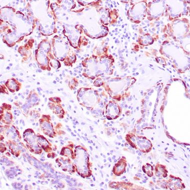
| Specificity | Positive control: Uterus, colon carcinoma |
Additional Info
| Tissue Specificity | High-molecular-weight caldesmon (isoform 1) is predominantly expressed in smooth muscles, whereas low-molecular-weight caldesmon (isoforms 2, 3, 4 and 5) are widely distributed in non-muscle tissues and cells. Not expressed in skeletal muscle or heart. |
| Post Translational Modifications | In non-muscle cells, phosphorylation by CDK1 during mitosis causes caldesmon to dissociate from microfilaments. Phosphorylation reduces caldesmon binding to actin, myosin, and calmodulin as well as its inhibition of actomyosin ATPase activity. Phosphorylation also occurs in both quiescent and dividing smooth muscle cells with similar effects on the interaction with actin and calmodulin and on microfilaments reorganization. CDK1-mediated phosphorylation promotes Schwann cell migration during peripheral nerve regeneration. |
| Function | Actin- and myosin-binding protein implicated in the regulation of actomyosin interactions in smooth muscle and nonmuscle cells (could act as a bridge between myosin and actin filaments). Stimulates actin binding of tropomyosin which increases the stabilization of actin filament structure. In muscle tissues, inhibits the actomyosin ATPase by binding to F-actin. This inhibition is attenuated by calcium-calmodulin and is potentiated by tropomyosin. Interacts with actin, myosin, two molecules of tropomyosin and with calmodulin. Also plays an essential role during cellular mitosis and receptor capping. Involved in Schwann cell migration during peripheral nerve regeneration. |
| Protein Name | CaldesmonCdm |
| Database Links | Reactome: R-HSA-445355 |
| Cellular Localisation | CytoplasmCytoskeletonMyofibrilStress FiberOn Thin Filaments In Smooth Muscle And On Stress Fibers In Fibroblasts (Nonmuscle) |
| Alternative Antibody Names | Anti-Caldesmon antibodyAnti-Cdm antibodyAnti-CALD1 antibodyAnti-CAD antibodyAnti-CDM antibody |
Information sourced from Uniprot.org