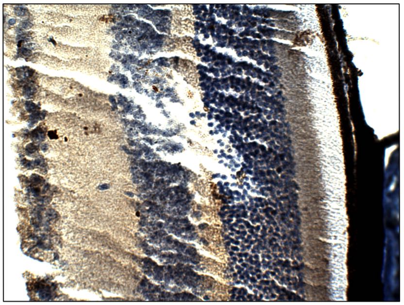
 at 1:50 dilution in Buffer. Section was treated with DAB and Haematoxylin stain. Magnification is at 40X.")
 at 1:50 dilution in Buffer. Section was treated with DAB and Haematoxylin stain. Magnification is at 40X.")
Anti-BEST1 antibody (Mid-region) (STJ500232)
SPECIFICATIONS
ClonalityPolyclonal
HostRabbit
ConjugationUnconjugated
IsotypeIgG
ImmunogenMid-Region Synthetic peptide corresponding to unique amino acid sequences on human Bestrophin 1 protein.
General Information
| Short Description | Rabbit polyclonal antibody anti-BEST1 (Mid-region) is suitable for use in Confocal Microscopy, ELISA, Immunofluorescence, Immunohistochemistry, Immunoprecipitation and Western Blot research applications. |
| Applications | CM/ELISA/IF/IHC/IP/WB |
| Host | Rabbit |
| Reactivity | Human/Monkey/Rat |
| Note | STRICTLY FOR FURTHER SCIENTIFIC RESEARCH USE ONLY (RUO). MUST NOT TO BE USED IN DIAGNOSTIC OR THERAPEUTIC APPLICATIONS. |
Product Properties
| Clonality | Polyclonal |
| Isotype | IgG |
| Conjugation | Unconjugated |
| Concentration | 0.65 µg/µl |
| Purification | Affinity Purified |
| Dilution Range | WB: 1:500DB: 1:10, 000ELISA: 1:10, 000IP: 1:200IHC: 1:100ICC: 1:100IF: 1:100CFM: 1:100 |
| Formulation | Contains Tris, HCl/Glycine buffer pH 7.4-7.8, 30% Glycerol and 0.5% BSA, along with cryo-protective agents, Hepes, and long-term preservatives (0.02% Sodium Azide). |
| Storage Instruction | Store at-20°C for long term storage. Avoid freeze-thaw cycles. |
Target Information
| Gene Symbol | BEST1 |
| Gene ID | 7439 |
| Uniprot ID | BEST1_HUMAN |
| Immunogen | Mid-Region Synthetic peptide corresponding to unique amino acid sequences on human Bestrophin 1 protein. |
| Immunogen Region | Mid-region |
Additional Info
| Tissue Specificity | Predominantly expressed in the basolateral membrane of the retinal pigment epithelium. |
| Function | Ligand-gated anion channel that allows the movement of anions across cell membranes when activated by calcium (Ca2+). Allows the movement of chloride and hydrogencarbonate. Found in a partially open conformation leading to significantly smaller chloride movement. Upon F2R/PAR-1 activation, the sequestered calcium is released into the cytosol of astrocytes, leading to the (Ca2+)-dependent release of L-glutamate into the synaptic cleft that targets the neuronal postsynaptic GRIN2A/NMDAR receptor resulting in the synaptic plasticity regulation. Upon activation of the norepinephrine-alpha-1 adrenergic receptor signaling pathway, transports as well D-serine than L-glutamate in a (Ca2+)-dependent manner, leading to activation of adjacent NMDAR receptors and therefore regulates the heterosynaptic long-term depression and metaplasticity during initial memory acquisition. Releases the 4-aminobutanoate neurotransmitter in a (Ca2+)-dependent manner, and participates in its tonic release from cerebellar glial cells. |
| Protein Name | Bestrophin-1Tu15bVitelliform Macular Dystrophy Protein 2 |
| Database Links | Reactome: R-HSA-2672351 |
| Cellular Localisation | Cell MembraneMulti-Pass Membrane ProteinBasolateral Cell MembraneLocalized At The Surface Membrane Of Microdomains Adjacent To Glutamatergic Synapses |
| Alternative Antibody Names | Anti-Bestrophin-1 antibodyAnti-Tu15b antibodyAnti-Vitelliform Macular Dystrophy Protein 2 antibodyAnti-BEST1 antibodyAnti-VMD2 antibody |
Information sourced from Uniprot.org