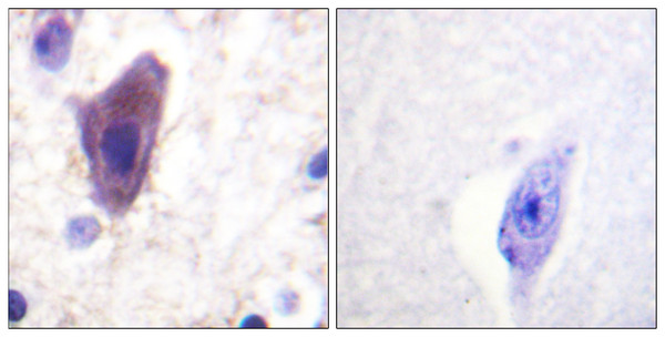
| Host: | Rabbit |
| Applications: | WB/IHC/IF/ELISA |
| Reactivity: | Human/Mouse |
| Note: | STRICTLY FOR FURTHER SCIENTIFIC RESEARCH USE ONLY (RUO). MUST NOT TO BE USED IN DIAGNOSTIC OR THERAPEUTIC APPLICATIONS. |
| Short Description : | Rabbit polyclonal antibody anti-Transferrin receptor protein 1 (15-64 aa) is suitable for use in Western Blot, Immunohistochemistry, Immunofluorescence and ELISA research applications. |
| Clonality : | Polyclonal |
| Conjugation: | Unconjugated |
| Isotype: | IgG |
| Formulation: | Liquid in PBS containing 50% Glycerol, 0.5% BSA and 0.02% Sodium Azide. |
| Purification: | The antibody was affinity-purified from rabbit antiserum by affinity-chromatography using epitope-specific immunogen. |
| Concentration: | 1 mg/mL |
| Dilution Range: | WB 1:500-1:2000IHC 1:100-1:300ELISA 1:10000IF 1:50-200 |
| Storage Instruction: | Store at-20°C for up to 1 year from the date of receipt, and avoid repeat freeze-thaw cycles. |
| Gene Symbol: | TFRC |
| Gene ID: | 7037 |
| Uniprot ID: | TFR1_HUMAN |
| Immunogen Region: | 15-64 aa |
| Specificity: | CD71 Polyclonal Antibody detects endogenous levels of CD71 protein. |
| Immunogen: | The antiserum was produced against synthesized peptide derived from the human CD71/TfR at the amino acid range 15-64 |
| Function | Cellular uptake of iron occurs via receptor-mediated endocytosis of ligand-occupied transferrin receptor into specialized endosomes. Endosomal acidification leads to iron release. The apotransferrin-receptor complex is then recycled to the cell surface with a return to neutral pH and the concomitant loss of affinity of apotransferrin for its receptor. Transferrin receptor is necessary for development of erythrocytes and the nervous system. A second ligand, the hereditary hemochromatosis protein HFE, competes for binding with transferrin for an overlapping C-terminal binding site. Positively regulates T and B cell proliferation through iron uptake. Acts as a lipid sensor that regulates mitochondrial fusion by regulating activation of the JNK pathway. When dietary levels of stearate (C18:0) are low, promotes activation of the JNK pathway, resulting in HUWE1-mediated ubiquitination and subsequent degradation of the mitofusin MFN2 and inhibition of mitochondrial fusion. When dietary levels of stearate (C18:0) are high, TFRC stearoylation inhibits activation of the JNK pathway and thus degradation of the mitofusin MFN2. Mediates uptake of NICOL1 into fibroblasts where it may regulate extracellular matrix production. (Microbial infection) Acts as a receptor for new-world arenaviruses: Guanarito, Junin and Machupo virus. |
| Protein Name | Transferrin Receptor Protein 1TrTfrTfr1TrfrT9P90Cd Antigen Cd71 Cleaved Into - Transferrin Receptor Protein 1 - Serum FormStfr |
| Database Links | Reactome: R-HSA-432722Reactome: R-HSA-8856825Reactome: R-HSA-8856828Reactome: R-HSA-8980692Reactome: R-HSA-9013026Reactome: R-HSA-9013106Reactome: R-HSA-9013148Reactome: R-HSA-9013149Reactome: R-HSA-9013404Reactome: R-HSA-9013406Reactome: R-HSA-9013407Reactome: R-HSA-9013408Reactome: R-HSA-9013409Reactome: R-HSA-9013423Reactome: R-HSA-917977Reactome: R-HSA-9696270Reactome: R-HSA-9696273Reactome: R-HSA-9725554 |
| Cellular Localisation | Cell MembraneSingle-Pass Type Ii Membrane ProteinMelanosomeIdentified By Mass Spectrometry In Melanosome Fractions From Stage I To Stage IvTransferrin Receptor Protein 1Serum Form: Secreted |
| Alternative Antibody Names | Anti-Transferrin Receptor Protein 1 antibodyAnti-Tr antibodyAnti-Tfr antibodyAnti-Tfr1 antibodyAnti-Trfr antibodyAnti-T9 antibodyAnti-P90 antibodyAnti-Cd Antigen Cd71 Cleaved Into - Transferrin Receptor Protein 1 - Serum Form antibodyAnti-Stfr antibodyAnti-TFRC antibody |
Information sourced from Uniprot.org


