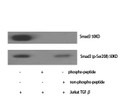
• Immunohistochemical analysis of paraffin-embedded Rat-kidney tissue. 1, Smad3 Polyclonal Antibody was diluted at 1:200 (4°C, overnight). 2, Sodium citrate pH 6.0 was used for antibody retrieval (>98°C, 20min). 3, Secondary antibody was diluted at 1:200 (room tempeRature, 30min). Negative control was used by secondary antibody only.
  • Immunohistochemical analysis of paraffin-embedded Rat-brain tissue. 1, Smad3 Polyclonal Antibody was diluted at 1:200 (4°C, overnight). 2, Sodium citrate pH 6.0 was used for antibody retrieval (>98°C, 20min). 3, Secondary antibody was diluted at 1:200 (room tempeRature, 30min). Negative control was used by secondary antibody only.
  • Immunofluorescence analysis of rat-lung tissue. 1, Smad3 Polyclonal Antibody (red) was diluted at 1:200 (4°C, overnight). 2, Cy3 labled Secondary antibody was diluted at 1:300 (room temperature, 50min).3, Picture B: DAPI (blue) 10min. Picture A:Target. Picture B: DAPI. Picture C: merge of A+B
  • Immunohistochemical analysis of paraffin-embedded Rat-testis tissue. 1, Smad3 Polyclonal Antibody was diluted at 1:200 (4°C, overnight). 2, Sodium citrate pH 6.0 was used for antibody retrieval (>98°C, 20min). 3, Secondary antibody was diluted at 1:200 (room tempeRature, 30min). Negative control was used by secondary antibody only.
  • Immunohistochemical analysis of paraffin-embedded Mouse-kidney tissue. 1, Smad3 Polyclonal Antibody was diluted at 1:200 (4°C, overnight). 2, Sodium citrate pH 6.0 was used for antibody retrieval (>98°C, 20min). 3, Secondary antibody was diluted at 1:200 (room tempeRature, 30min). Negative control was used by secondary antibody only.
  • Western blot analysis of HT29 cells using Smad3 Polyclonal Antibody diluted at 1:500
  • Immunohistochemical analysis of paraffin-embedded Human-uterus tissue. 1, Smad3 Polyclonal Antibody was diluted at 1:200 (4°C, overnight). 2, Sodium citrate pH 6.0 was used for antibody retrieval (>98°C, 20min). 3, Secondary antibody was diluted at 1:200 (room tempeRature, 30min). Negative control was used by secondary antibody only.
  • Immunohistochemical analysis of paraffin-embedded Mouse-colon tissue. 1, Smad3 Polyclonal Antibody was diluted at 1:200 (4°C, overnight). 2, Sodium citrate pH 6.0 was used for antibody retrieval (>98°C, 20min). 3, Secondary antibody was diluted at 1:200 (room tempeRature, 30min). Negative control was used by secondary antibody only.
  • Immunofluorescence analysis of mouse-kidney tissue. 1, Smad3 Polyclonal Antibody (red) was diluted at 1:200 (4°C, overnight). 2, Cy3 labled Secondary antibody was diluted at 1:300 (room temperature, 50min).3, Picture B: DAPI (blue) 10min. Picture A:Target. Picture B: DAPI. Picture C: merge of A+B
  • Immunofluorescence analysis of mouse-kidney tissue. 1, Smad3 Polyclonal Antibody (red) was diluted at 1:200 (4°C, overnight). 2, Cy3 labled Secondary antibody was diluted at 1:300 (room temperature, 50min).3, Picture B: DAPI (blue) 10min. Picture A:Target. Picture B: DAPI. Picture C: merge of A+B
  • Immunofluorescence analysis of rat-kidney tissue. 1, Smad3 Polyclonal Antibody (red) was diluted at 1:200 (4°C, overnight). 2, Cy3 labled Secondary antibody was diluted at 1:300 (room temperature, 50min).3, Picture B: DAPI (blue) 10min. Picture A:Target. Picture B: DAPI. Picture C: merge of A+B
  • Immunofluorescence analysis of rat-kidney tissue. 1, Smad3 Polyclonal Antibody (red) was diluted at 1:200 (4°C, overnight). 2, Cy3 labled Secondary antibody was diluted at 1:300 (room temperature, 50min).3, Picture B: DAPI (blue) 10min. Picture A:Target. Picture B: DAPI. Picture C: merge of A+B
  • Immunofluorescence analysis of rat-lung tissue. 1, Smad3 Polyclonal Antibody (red) was diluted at 1:200 (4°C, overnight). 2, Cy3 labled Secondary antibody was diluted at 1:300 (room temperature, 50min).3, Picture B: DAPI (blue) 10min. Picture A:Target. Picture B: DAPI. Picture C: merge of A+B
  • Immunohistochemical analysis of paraffin-embedded Mouse-heart tissue. 1, Smad3 Polyclonal Antibody was diluted at 1:200 (4°C, overnight). 2, Sodium citrate pH 6.0 was used for antibody retrieval (>98°C, 20min). 3, Secondary antibody was diluted at 1:200 (room tempeRature, 30min). Negative control was used by secondary antibody only.
  • Immunohistochemical analysis of paraffin-embedded Human-uterus-cancer tissue. 1, Smad3 Polyclonal Antibody was diluted at 1:200 (4°C, overnight). 2, Sodium citrate pH 6.0 was used for antibody retrieval (>98°C, 20min). 3, Secondary antibody was diluted at 1:200 (room tempeRature, 30min). Negative control was used by secondary antibody only.
  • Immunohistochemical analysis of paraffin-embedded Mouse-testis tissue. 1, Smad3 Polyclonal Antibody was diluted at 1:200 (4°C, overnight). 2, Sodium citrate pH 6.0 was used for antibody retrieval (>98°C, 20min). 3, Secondary antibody was diluted at 1:200 (room tempeRature, 30min). Negative control was used by secondary antibody only.
  • Immunohistochemical analysis of paraffin-embedded Human-colon tissue. 1, Smad3 Polyclonal Antibody was diluted at 1:200 (4°C, overnight). 2, Sodium citrate pH 6.0 was used for antibody retrieval (>98°C, 20min). 3, Secondary antibody was diluted at 1:200 (room tempeRature, 30min). Negative control was used by secondary antibody only.
  • Immunohistochemical analysis of paraffin-embedded Mouse-liver tissue. 1, Smad3 Polyclonal Antibody was diluted at 1:200 (4°C, overnight). 2, Sodium citrate pH 6.0 was used for antibody retrieval (>98°C, 20min). 3, Secondary antibody was diluted at 1:200 (room tempeRature, 30min). Negative control was used by secondary antibody only.
  • Immunohistochemical analysis of paraffin-embedded Human-lung tissue. 1, Smad3 Polyclonal Antibody was diluted at 1:200 (4°C, overnight). 2, Sodium citrate pH 6.0 was used for antibody retrieval (>98°C, 20min). 3, Secondary antibody was diluted at 1:200 (room tempeRature, 30min). Negative control was used by secondary antibody only.
  • Western blot analysis of various cells using Smad3 Polyclonal Antibody diluted at 1:500
  • Immunohistochemical analysis of paraffin-embedded Human-lung-cancer tissue. 1, Smad3 Polyclonal Antibody was diluted at 1:200 (4°C, overnight). 2, Sodium citrate pH 6.0 was used for antibody retrieval (>98°C, 20min). 3, Secondary antibody was diluted at 1:200 (room tempeRature, 30min). Negative control was used by secondary antibody only.
  • Western blot analysis of lysates from HT-29 cells, using Smad3 Antibody. The lane on the right is blocked with the synthesized peptide.
  • Immunofluorescence analysis of Hela cell. 1, Smad3 Polyclonal Antibody (red) was diluted at 1:200 (4°C overnight). Beta-actin monoclonal antibody (5B7) (green) was diluted at 1:200 (4°C overnight). 2, Goat Anti Rabbit Alexa Fluor 594 Catalog: (NA was diluted at 1:1000 (room temperature, 50min). Goat Anti Mouse Alexa Fluor 488 Catalog: (NA was diluted at 1:1000 (room temperature, 50min).

Anti-SMAD3 antibody (145-194 aa) (STJ95699)

SKU:
STJ95699

Current Stock:
Host: Rabbit
Applications: IF/WB/IHC/ELISA
Reactivity: Human/Mouse/Rat
Note: STRICTLY FOR FURTHER SCIENTIFIC RESEARCH USE ONLY (RUO). MUST NOT TO BE USED IN DIAGNOSTIC OR THERAPEUTIC APPLICATIONS.
Short Description: Rabbit polyclonal antibody anti-Mothers against decapentaplegic homolog 3 (145-194 aa) is suitable for use in Immunofluorescence, Western Blot, Immunohistochemistry and ELISA research applications.
Clonality: Polyclonal
Conjugation: Unconjugated
Isotype: IgG
Formulation: Liquid in PBS containing 50% Glycerol, 0.5% BSA and 0.02% Sodium Azide.
Purification: The antibody was affinity-purified from rabbit antiserum by affinity-chromatography using epitope-specific immunogen.
Concentration: 1 mg/mL
Dilution Range: IF 1:50-200
WB 1:500-1:2000
IHC 1:100-1:300
ELISA 1:10000
Storage Instruction: Store at-20°C for up to 1 year from the date of receipt, and avoid repeat freeze-thaw cycles.
Gene Symbol: SMAD3
Gene ID: 4088
Uniprot ID: SMAD3_HUMAN
Immunogen Region: 145-194 aa
Specificity: Smad3 Polyclonal Antibody detects endogenous levels of Smad3 protein.
Immunogen: The antiserum was produced against synthesized peptide derived from the human Smad3 at the amino acid range 145-194
Function Receptor-regulated SMAD (R-SMAD) that is an intracellular signal transducer and transcriptional modulator activated by TGF-beta (transforming growth factor) and activin type 1 receptor kinases. Binds the TRE element in the promoter region of many genes that are regulated by TGF-beta and, on formation of the SMAD3/SMAD4 complex, activates transcription. Also can form a SMAD3/SMAD4/JUN/FOS complex at the AP-1/SMAD site to regulate TGF-beta-mediated transcription. Has an inhibitory effect on wound healing probably by modulating both growth and migration of primary keratinocytes and by altering the TGF-mediated chemotaxis of monocytes. This effect on wound healing appears to be hormone-sensitive. Regulator of chondrogenesis and osteogenesis and inhibits early healing of bone fractures. Positively regulates PDPK1 kinase activity by stimulating its dissociation from the 14-3-3 protein YWHAQ which acts as a negative regulator.
Protein Name Mothers Against Decapentaplegic Homolog 3
Mad Homolog 3
Mad3
Mothers Against Dpp Homolog 3
Hmad-3
Jv15-2
Smad Family Member 3
Smad 3
Smad3
Hsmad3
Database Links Reactome: R-HSA-1181150
Reactome: R-HSA-1502540
Reactome: R-HSA-2173788
Reactome: R-HSA-2173789
Reactome: R-HSA-2173795
Reactome: R-HSA-2173796
Reactome: R-HSA-3304356
Reactome: R-HSA-3311021
Reactome: R-HSA-3315487
Reactome: R-HSA-3656532
Reactome: R-HSA-5689880
Reactome: R-HSA-8941855
Reactome: R-HSA-8952158
Reactome: R-HSA-9008059
Reactome: R-HSA-9013695
Reactome: R-HSA-9615017
Reactome: R-HSA-9617828
Reactome: R-HSA-9735871
Reactome: R-HSA-9754189
Reactome: R-HSA-9796292
Reactome: R-HSA-9823730
Reactome: R-HSA-9839394
Cellular Localisation Cytoplasm
Nucleus
Cytoplasmic And Nuclear In The Absence Of Tgf-Beta
On Tgf-Beta Stimulation
Migrates To The Nucleus When Complexed With Smad4
Through The Action Of The Phosphatase Ppm1a
Released From The Smad2/Smad4 Complex
And Exported Out Of The Nucleus By Interaction With Ranbp1
Co-Localizes With Lemd3 At The Nucleus Inner Membrane
Mapk-Mediated Phosphorylation Appears To Have No Effect On Nuclear Import
Pdpk1 Prevents Its Nuclear Translocation In Response To Tgf-Beta
Localized Mainly To The Nucleus In The Early Stages Of Embryo Development With Expression Becoming Evident In The Cytoplasm Of The Inner Cell Mass At The Blastocyst Stage
Alternative Antibody Names Anti-Mothers Against Decapentaplegic Homolog 3 antibody
Anti-Mad Homolog 3 antibody
Anti-Mad3 antibody
Anti-Mothers Against Dpp Homolog 3 antibody
Anti-Hmad-3 antibody
Anti-Jv15-2 antibody
Anti-Smad Family Member 3 antibody
Anti-Smad 3 antibody
Anti-Smad3 antibody
Anti-Hsmad3 antibody
Anti-SMAD3 antibody
Anti-MADH3 antibody

Information sourced from Uniprot.org

12 months for antibodies. 6 months for ELISA Kits. Please see website T&Cs for further guidance