


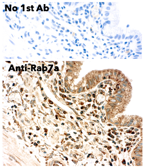
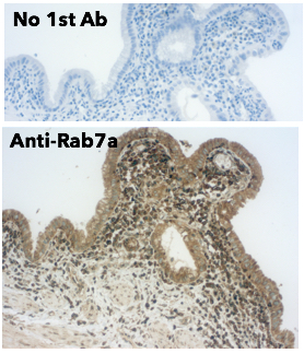
Anti-RAB7A antibody (C-Term) (STJ140063)
SPECIFICATIONS
ClonalityPolyclonal
HostGoat
ConjugationUnconjugated
IsotypeIgG
ImmunogenPurified recombinant peptide derived from within residues 115 aa to the C-terminus of mouse Rab5a produced in E. coli.
General Information
| Short Description | Goat polyclonal RAB7A, member RAS oncogene family (C-Term) antibody for use in WB, IF, IHC-P and IHC-F in human, mouse, rat, bovine, canine, chicken, avian, donkey, feline, goat, guinea pig, hamster, horse, porcine, rabbit, sheep and simian samples. |
| Applications | WB/IF/IHC-P/IHC-F |
| Host | Goat |
| Reactivity | Human/Mouse/Rat/Bovine/Canine/Chicken/Avian/Donkey/Feline/Goat/Guinea Pig/Hamster/Horse/Porcine/Rabbit/Sheep/Simian |
| Note | STRICTLY FOR FURTHER SCIENTIFIC RESEARCH USE ONLY (RUO). MUST NOT TO BE USED IN DIAGNOSTIC OR THERAPEUTIC APPLICATIONS. |
Product Properties
| Clonality | Polyclonal |
| Isotype | IgG |
| Conjugation | Unconjugated |
| Concentration | 3 mg/mL |
| Purification | This antibody is epitope-affinity purified from goat antiserum. |
| Dilution Range | WB 1:250-1:5000IF 1:50-1:250IHC-F 1:100-1:500IHC-P 1:100-1:500Cardoso MHS PhD Thesis NOVA University of Lisbon Portugal 2018 |
| Formulation | PBS, 20% Glycerol and 0.05% Sodium Azide. |
| Storage Instruction | For continuous use, store at 2-8 C for one-two days. For extended storage, store in-20 C freezer. Working dilution samples should be discarded if not used within 12 hours. |
Target Information
| Gene Symbol | RAB7A |
| Gene ID | 7879 |
| Uniprot ID | RAB7A_HUMAN |
| Accession Number | ENSG00000075785 |
| Immunogen | Purified recombinant peptide derived from within residues 115 aa to the C-terminus of mouse Rab5a produced in E. coli. |
| Immunogen Region | C-Term |
| Immunogen Sequence | FLIQASPRDPENFPFVVLGN LIQASPRDPENFPFVVLGNK IQASPRDPENFPFVVLGNKI QASPRDPENFPFVVLGNKID ASPRDPENFPFVVLGNKIDL SPRDPENFPFVVLGNKIDLE |
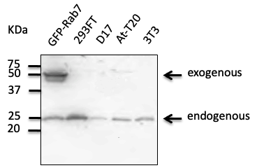
| Specificity | Detects Rab7a protein in the human, rat and mouse whole cell lysates and transfected cells with GFP-Rab7a by Western blot. This Ab is specific for Rab7a. |
Additional Info
| Tissue Specificity | Widely expressed.high expression found in skeletal muscle. |
| Post Translational Modifications | Deubiquitination at Lys-191 and Lys-194 by USP32. Phosphorylated at Ser-72 by LRRK1.phosphorylation is dependent on protein kinase C (PKC) activation of LRRK1. Prenylated. Prenylation is required for association with cellular membranes. |
| Function | The small GTPases Rab are key regulators of intracellular membrane trafficking, from the formation of transport vesicles to their fusion with membranes. Rabs cycle between an inactive GDP-bound form and an active GTP-bound form that is able to recruit to membranes different sets of downstream effectors directly responsible for vesicle formation, movement, tethering and fusion. In its active state, RAB7A binds to a variety of effector proteins playing a key role in the regulation of endo-lysosomal trafficking. Governs early-to-late endosomal maturation, microtubule minus-end as well as plus-end directed endosomal migration and positioning, and endosome-lysosome transport through different protein-protein interaction cascades. Also plays a central role in growth-factor-mediated cell signaling, nutrient-transportor mediated nutrient uptake, neurotrophin transport in the axons of neurons and lipid metabolism. Also involved in regulation of some specialized endosomal membrane trafficking, such as maturation of melanosomes, pathogen-induced phagosomes (or vacuoles) and autophagosomes. Plays a role in the maturation and acidification of phagosomes that engulf pathogens, such as S.aureus and M.tuberculosis. Plays a role in the fusion of phagosomes with lysosomes. In concert with RAC1, plays a role in regulating the formation of RBs (ruffled borders) in osteoclasts. Controls the endosomal trafficking and neurite outgrowth signaling of NTRK1/TRKA. Regulates the endocytic trafficking of the EGF-EGFR complex by regulating its lysosomal degradation. Involved in the ADRB2-stimulated lipolysis through lipophagy, a cytosolic lipase-independent autophagic pathway. Required for the exosomal release of SDCBP, CD63 and syndecan. Required for vesicular trafficking and cell surface expression of ACE2. May play a role in PRPH neuronal intermediate filament assembly. |
| Protein Name | Ras-Related Protein Rab-7a |
| Database Links | Reactome: R-HSA-2132295Reactome: R-HSA-6798695Reactome: R-HSA-8854214Reactome: R-HSA-8873719Reactome: R-HSA-8876198Reactome: R-HSA-9013148Reactome: R-HSA-9013149Reactome: R-HSA-9013404Reactome: R-HSA-9013405Reactome: R-HSA-9013406Reactome: R-HSA-9013407Reactome: R-HSA-9013408Reactome: R-HSA-9013409Reactome: R-HSA-9013423Reactome: R-HSA-9035034Reactome: R-HSA-9636383Reactome: R-HSA-9636569 |
| Cellular Localisation | Cytoplasmic VesiclePhagosome MembranePeripheral Membrane ProteinCytoplasmic SideLate Endosome MembraneLysosome MembraneMelanosome MembraneAutophagosome MembraneLipid DropletEndosome MembraneMitochondrion MembraneColocalizes With Osbpl1a At The Late EndosomeFound In The Ruffled Border (A Late Endosomal-Like Compartment In The Plasma Membrane) Of Bone-Resorbing OsteoclastsRecruited To Phagosomes Containing SAureus Or MycobacteriumLipid Droplet Localization Is Increased Upon Adrb2 StimulationRecruited To Damaged Mitochondria During Mitophagy In A Rimoc1-Dependent Manner |
| Alternative Antibody Names | Anti-Ras-Related Protein Rab-7a antibodyAnti-RAB7A antibodyAnti-RAB7 antibody |
Information sourced from Uniprot.org