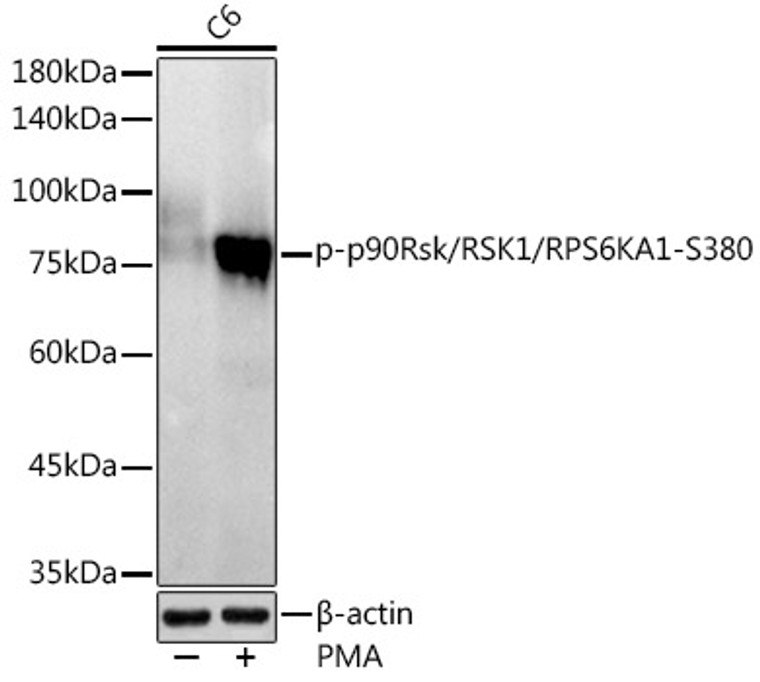
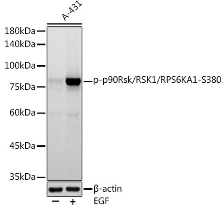
| Host: |
Rabbit |
| Applications: |
WB |
| Reactivity: |
Human/Mouse/Rat |
| Note: |
STRICTLY FOR FURTHER SCIENTIFIC RESEARCH USE ONLY (RUO). MUST NOT TO BE USED IN DIAGNOSTIC OR THERAPEUTIC APPLICATIONS. |
| Short Description: |
Rabbit monoclonal antibody anti-Phospho-RSK1-S380 is suitable for use in Western Blot research applications. |
| Clonality: |
Monoclonal |
| Clone ID: |
S1MR |
| Conjugation: |
Unconjugated |
| Isotype: |
IgG |
| Formulation: |
PBS with 0.02% Sodium Azide, 0.05% BSA, 50% Glycerol, pH7.3. |
| Purification: |
Affinity purification |
| Dilution Range: |
WB 1:500-1:1000 |
| Storage Instruction: |
Store at-20°C for up to 1 year from the date of receipt, and avoid repeat freeze-thaw cycles. |
| Gene Symbol: |
RPS6KA1 |
| Gene ID: |
6195 |
| Uniprot ID: |
KS6A1_HUMAN |
| Immunogen: |
A synthetic phosphorylated peptide around S380 of human p90Rsk/RSK1/RPS6KA1KS6A1 (Q15418). |
| Immunogen Sequence: |
GFSFV |
| Post Translational Modifications | Activated by phosphorylation at Ser-221 by PDPK1. Autophosphorylated on Ser-380, as part of the activation process. May be phosphorylated at Thr-359 and Ser-363 by MAPK1/ERK2 and MAPK3/ERK1. N-terminal myristoylation results in an activated kinase in the absence of added growth factors. |
| Function | Serine/threonine-protein kinase that acts downstream of ERK (MAPK1/ERK2 and MAPK3/ERK1) signaling and mediates mitogenic and stress-induced activation of the transcription factors CREB1, ETV1/ER81 and NR4A1/NUR77, regulates translation through RPS6 and EIF4B phosphorylation, and mediates cellular proliferation, survival, and differentiation by modulating mTOR signaling and repressing pro-apoptotic function of BAD and DAPK1. In fibroblast, is required for EGF-stimulated phosphorylation of CREB1, which results in the subsequent transcriptional activation of several immediate-early genes. In response to mitogenic stimulation (EGF and PMA), phosphorylates and activates NR4A1/NUR77 and ETV1/ER81 transcription factors and the cofactor CREBBP. Upon insulin-derived signal, acts indirectly on the transcription regulation of several genes by phosphorylating GSK3B at 'Ser-9' and inhibiting its activity. Phosphorylates RPS6 in response to serum or EGF via an mTOR-independent mechanism and promotes translation initiation by facilitating assembly of the pre-initiation complex. In response to insulin, phosphorylates EIF4B, enhancing EIF4B affinity for the EIF3 complex and stimulating cap-dependent translation. Is involved in the mTOR nutrient-sensing pathway by directly phosphorylating TSC2 at 'Ser-1798', which potently inhibits TSC2 ability to suppress mTOR signaling, and mediates phosphorylation of RPTOR, which regulates mTORC1 activity and may promote rapamycin-sensitive signaling independently of the PI3K/AKT pathway. Also involved in feedback regulation of mTORC1 and mTORC2 by phosphorylating DEPTOR. Mediates cell survival by phosphorylating the pro-apoptotic proteins BAD and DAPK1 and suppressing their pro-apoptotic function. Promotes the survival of hepatic stellate cells by phosphorylating CEBPB in response to the hepatotoxin carbon tetrachloride (CCl4). Mediates induction of hepatocyte prolifration by TGFA through phosphorylation of CEBPB. Is involved in cell cycle regulation by phosphorylating the CDK inhibitor CDKN1B, which promotes CDKN1B association with 14-3-3 proteins and prevents its translocation to the nucleus and inhibition of G1 progression. Phosphorylates EPHA2 at 'Ser-897', the RPS6KA-EPHA2 signaling pathway controls cell migration. In response to mTORC1 activation, phosphorylates EIF4B at 'Ser-406' and 'Ser-422' which stimulates bicarbonate cotransporter SLC4A7 mRNA translation, increasing SLC4A7 protein abundance and function. (Microbial infection) Promotes the late transcription and translation of viral lytic genes during Kaposi's sarcoma-associated herpesvirus/HHV-8 infection, when constitutively activated. |
| Protein Name | Ribosomal Protein S6 Kinase Alpha-1S6k-Alpha-190 Kda Ribosomal Protein S6 Kinase 1P90-Rsk 1P90rsk1P90s6kMap Kinase-Activated Protein Kinase 1aMapk-Activated Protein Kinase 1aMapkap Kinase 1aMapkapk-1aRibosomal S6 Kinase 1Rsk-1 |
| Database Links | Reactome: R-HSA-198753Reactome: R-HSA-199920Reactome: R-HSA-2559582Reactome: R-HSA-437239Reactome: R-HSA-442742Reactome: R-HSA-444257Reactome: R-HSA-881907 |
| Cellular Localisation | NucleusCytoplasm |
| Alternative Antibody Names | Anti-Ribosomal Protein S6 Kinase Alpha-1 antibodyAnti-S6k-Alpha-1 antibodyAnti-90 Kda Ribosomal Protein S6 Kinase 1 antibodyAnti-P90-Rsk 1 antibodyAnti-P90rsk1 antibodyAnti-P90s6k antibodyAnti-Map Kinase-Activated Protein Kinase 1a antibodyAnti-Mapk-Activated Protein Kinase 1a antibodyAnti-Mapkap Kinase 1a antibodyAnti-Mapkapk-1a antibodyAnti-Ribosomal S6 Kinase 1 antibodyAnti-Rsk-1 antibodyAnti-RPS6KA1 antibodyAnti-MAPKAPK1A antibodyAnti-RSK1 antibody |
Information sourced from Uniprot.org
12 months for antibodies. 6 months for ELISA Kits. Please see website T&Cs for further guidance


