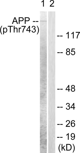
• Western blot analysis of lysates from HeLa cells, using Amyloid beta A4 (Phospho-Thr743/668) Antibody. The lane on the right is blocked with the phospho peptide.
  • Immunohistochemistry analysis of paraffin-embedded human brain, using Amyloid beta A4 (Phospho-Thr743/668) Antibody. The picture on the right is blocked with the phospho peptide.
  • Immunohistochemical analysis of paraffin-embedded Human breast cancer. Antibody was diluted at 1:100 (4°C overnight). High-pressure and temperature Tris-EDTA, pH8.0 was used for antigen retrieval. Negetive contrl (right) obtaned from antibody was pre-absorbed by immunogen peptide.
  • Immunofluorescence analysis of HeLa cells, using Amyloid beta A4 (Phospho-Thr743/668) Antibody. The picture on the right is blocked with the phospho peptide.
  • Western blot analysis of various cells using Phospho-Amyloid-Beta (T743) Polyclonal Antibody

Anti-Phospho-APP-Thr743 antibody (711-760 aa) (STJ91015)

SKU:
STJ91015

Current Stock:
Host: Rabbit
Applications: WB/IHC/IF/ELISA
Reactivity: Human/Mouse/Rat
Note: STRICTLY FOR FURTHER SCIENTIFIC RESEARCH USE ONLY (RUO). MUST NOT TO BE USED IN DIAGNOSTIC OR THERAPEUTIC APPLICATIONS.
Short Description: Rabbit polyclonal antibody anti-Phospho-Amyloid-beta precursor protein precursor protein-Thr743 (711-760 aa) is suitable for use in Western Blot, Immunohistochemistry, Immunofluorescence and ELISA research applications.
Clonality: Polyclonal
Conjugation: Unconjugated
Isotype: IgG
Formulation: Liquid in PBS containing 50% Glycerol, 0.5% BSA and 0.02% Sodium Azide.
Purification: The antibody was affinity-purified from rabbit antiserum by affinity-chromatography using epitope-specific immunogen.
Concentration: 1 mg/mL
Dilution Range: WB 1:500-1:2000
IHC 1:100-1:300
IF 1:200-1:1000
ELISA 1:10000
Storage Instruction: Store at-20°C for up to 1 year from the date of receipt, and avoid repeat freeze-thaw cycles.
Gene Symbol: APP
Gene ID: 351
Uniprot ID: A4_HUMAN
Immunogen Region: 711-760 aa
Specificity: Phospho-Amyloid-Beta (T743) Polyclonal Antibody detects endogenous levels of Amyloid-Beta protein only when phosphorylated at T743.
Immunogen: The antiserum was produced against synthesized peptide derived from the human Amyloid beta A4 around the phosphorylation site of Thr743/668 at the amino acid range 711-760
Post Translational Modifications Proteolytically processed under normal cellular conditions. Cleavage either by alpha-secretase, beta-secretase or theta-secretase leads to generation and extracellular release of soluble APP peptides, S-APP-alpha and S-APP-beta, and the retention of corresponding membrane-anchored C-terminal fragments, C80, C83 and C99. Subsequent processing of C80 and C83 by gamma-secretase yields P3 peptides. This is the major secretory pathway and is non-amyloidogenic. Alternatively, presenilin/nicastrin-mediated gamma-secretase processing of C99 releases the amyloid-beta proteins, amyloid-beta protein 40 and amyloid-beta protein 42, major components of amyloid plaques, and the cytotoxic C-terminal fragments, gamma-CTF(50), gamma-CTF(57) and gamma-CTF(59). PSEN1 cleavage is more efficient with C83 than with C99 as substrate (in vitro). Amyloid-beta protein 40 and Amyloid-beta protein 42 are cleaved by ACE. Many other minor amyloid-beta peptides, amyloid-beta 1-X peptides, are found in cerebral spinal fluid (CSF) including the amyloid-beta X-15 peptides, produced from the cleavage by alpha-secretase and all terminating at Gln-686. Proteolytically cleaved by caspases during neuronal apoptosis. Cleavage at Asp-739 by either CASP6, CASP8 or CASP9 results in the production of the neurotoxic C31 peptide and the increased production of amyloid-beta peptides. N-glycosylated. N- and O-glycosylated. O-glycosylation on Ser and Thr residues with core 1 or possibly core 8 glycans. Partial tyrosine glycosylation (Tyr-681) is found on some minor, short amyloid-beta peptides (amyloid-beta 1-15, 1-16, 1-17, 1-18, 1-19 and 1-20) but not found on amyloid-beta protein 38, amyloid-beta protein 40 nor on amyloid-beta protein 42. Modification on a tyrosine is unusual and is more prevelant in AD patients. Glycans had Neu5AcHex(Neu5Ac)HexNAc-O-Tyr, Neu5AcNeu5AcHex(Neu5Ac)HexNAc-O-Tyr and O-AcNeu5AcNeu5AcHex(Neu5Ac)HexNAc-O-Tyr structures, where O-Ac is O-acetylation of Neu5Ac. Neu5AcNeu5Ac is most likely Neu5Ac 2,8Neu5Ac linked. O-glycosylations in the vicinity of the cleavage sites may influence the proteolytic processing. Appicans are L-APP isoforms with O-linked chondroitin sulfate. Phosphorylation in the C-terminal on tyrosine, threonine and serine residues is neuron-specific. Phosphorylation can affect APP processing, neuronal differentiation and interaction with other proteins. Phosphorylated on Thr-743 in neuronal cells by Cdc5 kinase and Mapk10, in dividing cells by Cdc2 kinase in a cell-cycle dependent manner with maximal levels at the G2/M phase and, in vitro, by GSK-3-beta. The Thr-743 phosphorylated form causes a conformational change which reduces binding of Fe65 family members. In dopaminergic (DA) neurons, phosphorylation on Thr-743 by LRKK2 promotes the production and the nuclear translocation of the APP intracellular domain (AICD) which induces DA neuron apoptosis. Phosphorylation on Tyr-757 is required for SHC binding. Phosphorylated in the extracellular domain by casein kinases on both soluble and membrane-bound APP. This phosphorylation is inhibited by heparin. Extracellular binding and reduction of copper, results in a corresponding oxidation of Cys-144 and Cys-158, and the formation of a disulfide bond. In vitro, the APP-Cu(+) complex in the presence of hydrogen peroxide results in an increased production of amyloid-beta-containing peptides. Trophic-factor deprivation triggers the cleavage of surface APP by beta-secretase to release sAPP-beta which is further cleaved to release an N-terminal fragment of APP (N-APP). Amyloid-beta peptides are degraded by IDE. Sulfated on tyrosine residues.
Function Functions as a cell surface receptor and performs physiological functions on the surface of neurons relevant to neurite growth, neuronal adhesion and axonogenesis. Interaction between APP molecules on neighboring cells promotes synaptogenesis. Involved in cell mobility and transcription regulation through protein-protein interactions. Can promote transcription activation through binding to APBB1-KAT5 and inhibits Notch signaling through interaction with Numb. Couples to apoptosis-inducing pathways such as those mediated by G(o) and JIP. Inhibits G(o) alpha ATPase activity. Acts as a kinesin I membrane receptor, mediating the axonal transport of beta-secretase and presenilin 1. By acting as a kinesin I membrane receptor, plays a role in axonal anterograde transport of cargo towards synapses in axons. Involved in copper homeostasis/oxidative stress through copper ion reduction. In vitro, copper-metallated APP induces neuronal death directly or is potentiated through Cu(2+)-mediated low-density lipoprotein oxidation. Can regulate neurite outgrowth through binding to components of the extracellular matrix such as heparin and collagen I and IV. The splice isoforms that contain the BPTI domain possess protease inhibitor activity. Induces a AGER-dependent pathway that involves activation of p38 MAPK, resulting in internalization of amyloid-beta peptide and leading to mitochondrial dysfunction in cultured cortical neurons. Provides Cu(2+) ions for GPC1 which are required for release of nitric oxide (NO) and subsequent degradation of the heparan sulfate chains on GPC1. Amyloid-beta peptides are lipophilic metal chelators with metal-reducing activity. Bind transient metals such as copper, zinc and iron. In vitro, can reduce Cu(2+) and Fe(3+) to Cu(+) and Fe(2+), respectively. Amyloid-beta protein 42 is a more effective reductant than amyloid-beta protein 40. Amyloid-beta peptides bind to lipoproteins and apolipoproteins E and J in the CSF and to HDL particles in plasma, inhibiting metal-catalyzed oxidation of lipoproteins. APP42-beta may activate mononuclear phagocytes in the brain and elicit inflammatory responses. Promotes both tau aggregation and TPK II-mediated phosphorylation. Interaction with overexpressed HADH2 leads to oxidative stress and neurotoxicity. Also binds GPC1 in lipid rafts. Appicans elicit adhesion of neural cells to the extracellular matrix and may regulate neurite outgrowth in the brain. The gamma-CTF peptides as well as the caspase-cleaved peptides, including C31, are potent enhancers of neuronal apoptosis. N-APP binds TNFRSF21 triggering caspase activation and degeneration of both neuronal cell bodies (via caspase-3) and axons (via caspase-6).
Protein Name Amyloid-Beta Precursor Protein
App
Abpp
Appi
Alzheimer Disease Amyloid A4 Protein Homolog
Alzheimer Disease Amyloid Protein
Amyloid Precursor Protein
Amyloid-Beta
A4 Precursor Protein
Amyloid-Beta A4 Protein
Cerebral Vascular Amyloid Peptide
Cvap
Prea4
Protease Nexin-Ii
Pn-Ii Cleaved Into - N-App - Soluble App-Alpha
S-App-Alpha - Soluble App-Beta
S-App-Beta - C99
Beta-Secretase C-Terminal Fragment
Beta-Ctf - Amyloid-Beta Protein 42
Abeta42
Beta-App42 - Amyloid-Beta Protein 40
Abeta40
Beta-App40 - C83
Alpha-Secretase C-Terminal Fragment
Alpha-Ctf - P3(42 - P3(40 - C80 - Gamma-Secretase C-Terminal Fragment 59
Amyloid Intracellular Domain 59
Aicd-59
Aid(59
Gamma-Ctf(59 - Gamma-Secretase C-Terminal Fragment 57
Amyloid Intracellular Domain 57
Aicd-57
Aid(57
Gamma-Ctf(57 - Gamma-Secretase C-Terminal Fragment 50
Amyloid Intracellular Domain 50
Aicd-50
Aid(50
Gamma-Ctf(50 - C31
Database Links Reactome: R-HSA-114608
Reactome: R-HSA-3000178
Reactome: R-HSA-381426
Reactome: R-HSA-416476
Reactome: R-HSA-418594
Reactome: R-HSA-432720
Reactome: R-HSA-444473
Reactome: R-HSA-445989
Reactome: R-HSA-844456
Reactome: R-HSA-879415
Reactome: R-HSA-8862803
Reactome: R-HSA-8957275
Reactome: R-HSA-933542
Reactome: R-HSA-9609523
Reactome: R-HSA-9660826
Reactome: R-HSA-977225
Cellular Localisation Cell Membrane
Single-Pass Type I Membrane Protein
Membrane
Perikaryon
Cell Projection
Growth Cone
Clathrin-Coated Pit
Early Endosome
Cytoplasmic Vesicle
Cell Surface Protein That Rapidly Becomes Internalized Via Clathrin-Coated Pits
Only A Minor Proportion Is Present At The Cell Membrane
Most Of The Protein Is Present In Intracellular Vesicles
During Maturation
The Immature App (N-Glycosylated In The Endoplasmic Reticulum) Moves To The Golgi Complex Where Complete Maturation Occurs (O-Glycosylated And Sulfated)
After Alpha-Secretase Cleavage
Soluble App Is Released Into The Extracellular Space And The C-Terminal Is Internalized To Endosomes And Lysosomes
Some App Accumulates In Secretory Transport Vesicles Leaving The Late Golgi Compartment And Returns To The Cell Surface
App Sorts To The Basolateral Surface In Epithelial Cells
During Neuronal Differentiation
The Thr-743 Phosphorylated Form Is Located Mainly In Growth Cones
Moderately In Neurites And Sparingly In The Cell Body
Casein Kinase Phosphorylation Can Occur Either At The Cell Surface Or Within A Post-Golgi Compartment
Associates With Gpc1 In Perinuclear Compartments
Colocalizes With Sorl1 In A Vesicular Pattern In Cytoplasm And Perinuclear Regions
C83: Endoplasmic Reticulum
Golgi Apparatus
C99: Early Endosome
Soluble App-Beta: Secreted
Amyloid-Beta Protein 40: Cell Surface
Amyloid-Beta Protein 42: Cell Surface
Associates With Fpr2 At The Cell Surface And The Complex Is Then Rapidly Internalized
Gamma-Secretase C-Terminal Fragment 59: Nucleus
Cytoplasm
Located To Both The Cytoplasm And Nuclei Of Neurons
It Can Be Translocated To The Nucleus Through Association With Apbb1 (Fe65)
In Dopaminergic Neurons
The Phosphorylated Thr-743 Form Is Localized To The Nucleus
Alternative Antibody Names Anti-Amyloid-Beta Precursor Protein antibody
Anti-App antibody
Anti-Abpp antibody
Anti-Appi antibody
Anti-Alzheimer Disease Amyloid A4 Protein Homolog antibody
Anti-Alzheimer Disease Amyloid Protein antibody
Anti-Amyloid Precursor Protein antibody
Anti-Amyloid-Beta antibody
Anti-A4 Precursor Protein antibody
Anti-Amyloid-Beta A4 Protein antibody
Anti-Cerebral Vascular Amyloid Peptide antibody
Anti-Cvap antibody
Anti-Prea4 antibody
Anti-Protease Nexin-Ii antibody
Anti-Pn-Ii Cleaved Into - N-App - Soluble App-Alpha antibody
Anti-S-App-Alpha - Soluble App-Beta antibody
Anti-S-App-Beta - C99 antibody
Anti-Beta-Secretase C-Terminal Fragment antibody
Anti-Beta-Ctf - Amyloid-Beta Protein 42 antibody
Anti-Abeta42 antibody
Anti-Beta-App42 - Amyloid-Beta Protein 40 antibody
Anti-Abeta40 antibody
Anti-Beta-App40 - C83 antibody
Anti-Alpha-Secretase C-Terminal Fragment antibody
Anti-Alpha-Ctf - P3(42 - P3(40 - C80 - Gamma-Secretase C-Terminal Fragment 59 antibody
Anti-Amyloid Intracellular Domain 59 antibody
Anti-Aicd-59 antibody
Anti-Aid(59 antibody
Anti-Gamma-Ctf(59 - Gamma-Secretase C-Terminal Fragment 57 antibody
Anti-Amyloid Intracellular Domain 57 antibody
Anti-Aicd-57 antibody
Anti-Aid(57 antibody
Anti-Gamma-Ctf(57 - Gamma-Secretase C-Terminal Fragment 50 antibody
Anti-Amyloid Intracellular Domain 50 antibody
Anti-Aicd-50 antibody
Anti-Aid(50 antibody
Anti-Gamma-Ctf(50 - C31 antibody
Anti-APP antibody
Anti-A4 antibody
Anti-AD1 antibody

Information sourced from Uniprot.org

12 months for antibodies. 6 months for ELISA Kits. Please see website T&Cs for further guidance