| Post Translational Modifications | Phosphorylated by PRKCZ, which may prevent MutS alpha degradation by the ubiquitin-proteasome pathway. |
| Function | Component of the post-replicative DNA mismatch repair system (MMR). Forms two different heterodimers: MutS alpha (MSH2-MSH6 heterodimer) and MutS beta (MSH2-MSH3 heterodimer) which binds to DNA mismatches thereby initiating DNA repair. When bound, heterodimers bend the DNA helix and shields approximately 20 base pairs. MutS alpha recognizes single base mismatches and dinucleotide insertion-deletion loops (IDL) in the DNA. MutS beta recognizes larger insertion-deletion loops up to 13 nucleotides long. After mismatch binding, MutS alpha or beta forms a ternary complex with the MutL alpha heterodimer, which is thought to be responsible for directing the downstream MMR events, including strand discrimination, excision, and resynthesis. Recruits DNA helicase MCM9 to chromatin which unwinds the mismatch containing DNA strand. ATP binding and hydrolysis play a pivotal role in mismatch repair functions. The ATPase activity associated with MutS alpha regulates binding similar to a molecular switch: mismatched DNA provokes ADP-->ATP exchange, resulting in a discernible conformational transition that converts MutS alpha into a sliding clamp capable of hydrolysis-independent diffusion along the DNA backbone. This transition is crucial for mismatch repair. MutS alpha may also play a role in DNA homologous recombination repair. In melanocytes may modulate both UV-B-induced cell cycle regulation and apoptosis. |
| Protein Name | Dna Mismatch Repair Protein Msh2Hmsh2Muts Protein Homolog 2 |
| Database Links | Reactome: R-HSA-5358565Reactome: R-HSA-5358606Reactome: R-HSA-5632927Reactome: R-HSA-5632928Reactome: R-HSA-5632968Reactome: R-HSA-6796648 |
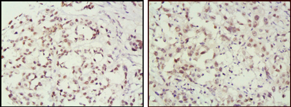
| Cellular Localisation | NucleusChromosome |
| Alternative Antibody Names | Anti-Dna Mismatch Repair Protein Msh2 antibodyAnti-Hmsh2 antibodyAnti-Muts Protein Homolog 2 antibodyAnti-MSH2 antibody |
Information sourced from Uniprot.org


