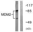
| Post Translational Modifications | Phosphorylation on Ser-166 by SGK1 activates ubiquitination of p53/TP53. Phosphorylated at multiple sites near the RING domain by ATM upon DNA damage.this promotes its ubiquitination and degradation, preventing p53/TP53 degradation. Autoubiquitination leads to proteasomal degradation.resulting in p53/TP53 activation it may be regulated by SFN. Also ubiquitinated by TRIM13. ATM-phosphorylated MDM2 is ubiquitinated by the SCF(FBXO31) complex in response to genotoxic stress, promoting its degradation and p53/TP53-mediated DNA damage response. Deubiquitinated by USP2 leads to its accumulation and increases deubiquitination and degradation of p53/TP53. Deubiquitinated by USP7 leading to its stabilization. |
| Function | E3 ubiquitin-protein ligase that mediates ubiquitination of p53/TP53, leading to its degradation by the proteasome. Inhibits p53/TP53- and p73/TP73-mediated cell cycle arrest and apoptosis by binding its transcriptional activation domain. Also acts as a ubiquitin ligase E3 toward itself and ARRB1. Permits the nuclear export of p53/TP53. Promotes proteasome-dependent ubiquitin-independent degradation of retinoblastoma RB1 protein. Inhibits DAXX-mediated apoptosis by inducing its ubiquitination and degradation. Component of the TRIM28/KAP1-MDM2-p53/TP53 complex involved in stabilizing p53/TP53. Also a component of the TRIM28/KAP1-ERBB4-MDM2 complex which links growth factor and DNA damage response pathways. Mediates ubiquitination and subsequent proteasome degradation of DYRK2 in nucleus. Ubiquitinates IGF1R and SNAI1 and promotes them to proteasomal degradation. Ubiquitinates DCX, leading to DCX degradation and reduction of the dendritic spine density of olfactory bulb granule cells. Ubiquitinates DLG4, leading to proteasomal degradation of DLG4 which is required for AMPA receptor endocytosis. Negatively regulates NDUFS1, leading to decreased mitochondrial respiration, marked oxidative stress, and commitment to the mitochondrial pathway of apoptosis. Binds NDUFS1 leading to its cytosolic retention rather than mitochondrial localization resulting in decreased supercomplex assembly (interactions between complex I and complex III), decreased complex I activity, ROS production, and apoptosis. |
| Protein Name | E3 Ubiquitin-Protein Ligase Mdm2Double Minute 2 ProteinHdm2Oncoprotein Mdm2Ring-Type E3 Ubiquitin Transferase Mdm2P53-Binding Protein Mdm2 |
| Database Links | Reactome: R-HSA-198323Reactome: R-HSA-2559580Reactome: R-HSA-2559585Reactome: R-HSA-3232118Reactome: R-HSA-3232142Reactome: R-HSA-399719Reactome: R-HSA-5674400Reactome: R-HSA-5689880Reactome: R-HSA-6804756Reactome: R-HSA-6804757Reactome: R-HSA-6804760Reactome: R-HSA-69541Reactome: R-HSA-8941858Reactome: R-HSA-9725370Reactome: R-HSA-9768919 |
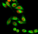
| Cellular Localisation | NucleusNucleoplasmCytoplasmNucleolusExpressed Predominantly In The NucleoplasmInteraction With Arf(P14) Results In The Localization Of Both Proteins To The NucleolusThe Nucleolar Localization Signals In Both Arf(P14) And Mdm2 May Be Necessary To Allow Efficient Nucleolar Localization Of Both ProteinsColocalizes With Rassf1 Isoform A In The Nucleus |
| Alternative Antibody Names | Anti-E3 Ubiquitin-Protein Ligase Mdm2 antibodyAnti-Double Minute 2 Protein antibodyAnti-Hdm2 antibodyAnti-Oncoprotein Mdm2 antibodyAnti-Ring-Type E3 Ubiquitin Transferase Mdm2 antibodyAnti-P53-Binding Protein Mdm2 antibodyAnti-MDM2 antibody |
Information sourced from Uniprot.org


