

. 2, STJ92908")

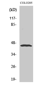
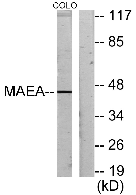
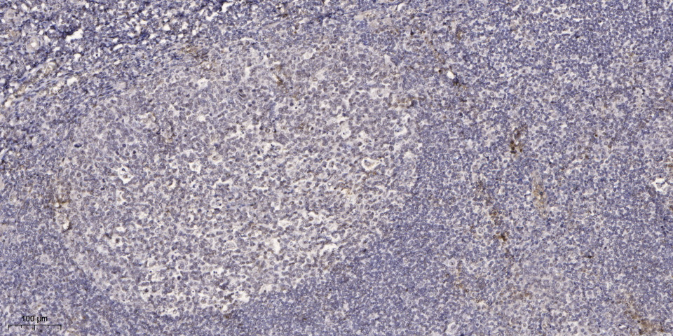
Anti-MAEA antibody (181-230 aa) (STJ92908)
SPECIFICATIONS
ClonalityPolyclonal
HostRabbit
ConjugationUnconjugated
IsotypeIgG
ImmunogenThe antiserum was produced against synthesized peptide derived from the human MAEA at the amino acid range 181-230
General Information
| Short Description | Rabbit polyclonal anti-E3 ubiquitin-protein transferase MAEA (181-230 aa) for use in WB, IHC, IF and ELISA in Human, Mouse and Rat samples. Datasheet included with dilution recommendations, and related reagents. |
| Applications | WB/IHC/IF/ELISA |
| Host | Rabbit |
| Reactivity | Human/Mouse/Rat |
| Note | STRICTLY FOR FURTHER SCIENTIFIC RESEARCH USE ONLY (RUO). MUST NOT TO BE USED IN DIAGNOSTIC OR THERAPEUTIC APPLICATIONS. |
Product Properties
| Clonality | Polyclonal |
| Isotype | IgG |
| Conjugation | Unconjugated |
| Concentration | 1 mg/mL |
| Purification | The antibody was affinity-purified from rabbit antiserum by affinity-chromatography using epitope-specific immunogen. |
| Dilution Range | WB 1:500-1:2000IHC 1:100-1:300ELISA 1:20000IF 1:50-200 |
| Formulation | Liquid in PBS containing 50% Glycerol, 0.5% BSA and 0.02% Sodium Azide. |
| Storage Instruction | Store at-20°C for up to 1 year from the date of receipt, and avoid repeat freeze-thaw cycles. |
Target Information
| Gene Symbol | MAEA |
| Gene ID | 10296 |
| Uniprot ID | MAEA_HUMAN |
| Immunogen | The antiserum was produced against synthesized peptide derived from the human MAEA at the amino acid range 181-230 |
| Immunogen Region | 181-230 aa |
| Specificity | Emp Polyclonal Antibody detects endogenous levels of Emp protein. |
Additional Info
| Post Translational Modifications | Autoubiquitinated as component of the CTLH E3 ubiquitin-protein ligase complex (in vitro). |
| Function | Core component of the CTLH E3 ubiquitin-protein ligase complex that selectively accepts ubiquitin from UBE2H and mediates ubiquitination and subsequent proteasomal degradation of the transcription factor HBP1. MAEA and RMND5A are both required for catalytic activity of the CTLH E3 ubiquitin-protein ligase complex. MAEA is required for normal cell proliferation. The CTLH E3 ubiquitin-protein ligase complex is not required for the degradation of enzymes involved in gluconeogenesis, such as FBP1. Plays a role in erythroblast enucleation during erythrocyte maturation and in the development of mature macrophages. Mediates the attachment of erythroid cell to mature macrophages.this MAEA-mediated contact inhibits erythroid cell apoptosis. Participates in erythroblastic island formation, which is the functional unit of definitive erythropoiesis. Associates with F-actin to regulate actin distribution in erythroblasts and macrophages. May contribute to nuclear architecture and cells division events (Probable). |
| Protein Name | E3 Ubiquitin-Protein Transferase MaeaCell Proliferation-Inducing Gene 5 ProteinErythroblast Macrophage ProteinHuman Lung Cancer Oncogene 10 ProteinHlc-10Macrophage Erythroblast AttacherP44emlp |
| Database Links | Reactome: R-HSA-9861718 |
| Cellular Localisation | CytoplasmNucleusNucleoplasmNucleus MatrixCell MembraneCytoskeletonDetected In A NuclearSpeckled-Like PatternLocalized With Condensed Chromatin At ProphaseDetected In Nuclear Spindle Poles At Metaphase And In The Contractile Ring During Telophase And CytokinesisPresent In CytoplasmNuclear Matrix And At The Cell Surface In MacrophagesPredominantly Nuclear In Immature Macrophages And Predominantly Detected At The Cell Surface In Mature MacrophagesColocalizes With F-Actin In Macrophages |
| Alternative Antibody Names | Anti-E3 Ubiquitin-Protein Transferase Maea antibodyAnti-Cell Proliferation-Inducing Gene 5 Protein antibodyAnti-Erythroblast Macrophage Protein antibodyAnti-Human Lung Cancer Oncogene 10 Protein antibodyAnti-Hlc-10 antibodyAnti-Macrophage Erythroblast Attacher antibodyAnti-P44emlp antibodyAnti-MAEA antibodyAnti-EMP antibodyAnti-HLC10 antibodyAnti-PIG5 antibody |
Information sourced from Uniprot.org