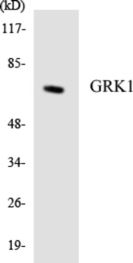
| Host: | Rabbit |
| Applications: | WB/IHC/IF/ELISA |
| Reactivity: | Human/Mouse/Rat |
| Note: | STRICTLY FOR FURTHER SCIENTIFIC RESEARCH USE ONLY (RUO). MUST NOT TO BE USED IN DIAGNOSTIC OR THERAPEUTIC APPLICATIONS. |
| Short Description : | Rabbit polyclonal anti-Rhodopsin kinase GRK1 (6-55 aa) for use in WB, IHC, IF and ELISA in Human, Mouse and Rat samples. Datasheet included with dilution recommendations, and related reagents. |
| Clonality : | Polyclonal |
| Conjugation: | Unconjugated |
| Isotype: | IgG |
| Formulation: | Liquid in PBS containing 50% Glycerol, 0.5% BSA and 0.02% Sodium Azide. |
| Purification: | The antibody was affinity-purified from rabbit antiserum by affinity-chromatography using epitope-specific immunogen. |
| Concentration: | 1 mg/mL |
| Dilution Range: | WB 1:500-1:2000IHC 1:100-1:300ELISA 1:40000IF 1:50-200 |
| Storage Instruction: | Store at-20°C for up to 1 year from the date of receipt, and avoid repeat freeze-thaw cycles. |
| Gene Symbol: | GRK1 |
| Gene ID: | 6011 |
| Uniprot ID: | GRK1_HUMAN |
| Immunogen Region: | 6-55 aa |
| Specificity: | GRK 1 Polyclonal Antibody detects endogenous levels of GRK 1 protein. |
| Immunogen: | The antiserum was produced against synthesized peptide derived from the human GRK1 at the amino acid range 6-55 |
| Post Translational Modifications | Autophosphorylated, Ser-21 is a minor site of autophosphorylation compared to Ser-491 and Thr-492. Phosphorylation at Ser-21 is regulated by light and activated by cAMP. Farnesylation is required for full activity. |
| Function | Retina-specific kinase involved in the signal turnoff via phosphorylation of rhodopsin (RHO), the G protein- coupled receptor that initiates the phototransduction cascade. This rapid desensitization is essential for scotopic vision and permits rapid adaptation to changes in illumination. May play a role in the maintenance of the outer nuclear layer in the retina. |
| Protein Name | Rhodopsin Kinase Grk1RkG Protein-Coupled Receptor Kinase 1 |
| Database Links | Reactome: R-HSA-2514859 |
| Cellular Localisation | MembraneLipid-AnchorCell ProjectionCiliumPhotoreceptor Outer SegmentSubcellular Location Is Not Affected By Light Or Dark Conditions |
| Alternative Antibody Names | Anti-Rhodopsin Kinase Grk1 antibodyAnti-Rk antibodyAnti-G Protein-Coupled Receptor Kinase 1 antibodyAnti-GRK1 antibodyAnti-RHOK antibody |
Information sourced from Uniprot.org


