


Anti-GFAP antibody (STJA0003659)
SPECIFICATIONS
ClonalityPolyclonal
HostChicken
ConjugationUnconjugated
IsotypeIgY
ImmunogenRecombinant and purification bovine GFAP.
General Information
| Short Description | Chicken polyclonal anti-GFAP for use in WB, IHC and ICC in Bovine, Human, Mouse and Rat samples. Datasheet included with dilution recommendations, and related reagents. |
| Applications | WB/IHC/ICC |
| Host | Chicken |
| Reactivity | Bovine/Human/Mouse/Rat |
| Note | STRICTLY FOR FURTHER SCIENTIFIC RESEARCH USE ONLY (RUO). MUST NOT TO BE USED IN DIAGNOSTIC OR THERAPEUTIC APPLICATIONS. |
Product Properties
| Clonality | Polyclonal |
| Isotype | IgY |
| Conjugation | Unconjugated |
| Purification | This antibody was total igy fraction. |
| Dilution Range | WB 1:10, 000IHC 1:1000-1:5000ICC 1:1000-1:5000 |
| Formulation | Total IgY fraction in PBS + 10 mM Sodium Azide. |
| Storage Instruction | Store at-20°C for up to 1 year from the date of receipt, and avoid repeat freeze-thaw cycles. |
Target Information
| Immunogen | Recombinant and purification bovine GFAP. |
Additional Info
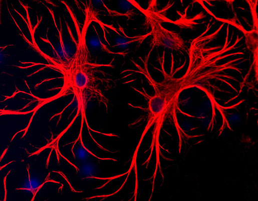
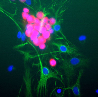
| Background | Glial Fibrillary Acidic Protein (GFAP) was discovered by Amico Bignami and co-workers as a major fibrous protein of multiple sclerosis plaques (Bignami at al., 1972). It was subsequently found to be a member of the 10nm or intermediate filament (IF) family, specifically the IF family Class III, which also includes peripherin, desmin and vimentin. GFAP is strongly and specifically expressed in astrocytes and certain other astroglia in the CNS, in satellite cells, peripheral ganglia, and in non-myelinating Schwann cells in peripheral nerves. In many damage and disease states GFAP expression is heavily upregulated in astrocytes. In addition, neural stem cells frequently strongly express GFAP. Point mutations in the protein coding region of the GFAP gene lead to Alexander disease which is characterized by the presence of abnormal astrocytes containing GFAP protein aggregates known as Rosenthal fibers (Brenner et al., 2001). Western blot of rat cortical lysate showing specific immunolabeling of the ~50 kDa GFAP protein. |
Information sourced from Uniprot.org