• Confocal imaging of paraffin-embedded Mouse colon tissue using ASH2L Rabbit monoclonal antibody (STJ11102184, dilution 1:100) followed by a further incubation with Cy3 Goat Anti-Rabbit IgG (H+L) ( dilution 1:500) (Red). DAPI was used for nuclear staining (Blue). Objective: 40x. Perform high pressure antigen retrieval with 0. 01 M citrate buffer (pH 6. 0) prior to IF staining.
  • Confocal imaging of NIH/3T3 cells using ASH2L Rabbit monoclonal antibody (STJ11102184, dilution 1:100) followed by a further incubation with Cy3 Goat Anti-Rabbit IgG (H+L) ( dilution 1:500) (Red). The cells were counterstained with Alpha-Tubulin Mouse monoclonal antibody ( dilution 1:400) followed by incubation with ABflo® 488-conjugated Goat Anti-Mouse IgG (H+L) antibody ( dilution 1:500) (Green). DAPI was used for nuclear staining (Blue). Objective: 100x.
  • Confocal imaging of C2C12 cells using ASH2L Rabbit monoclonal antibody (STJ11102184, dilution 1:100) followed by a further incubation with Cy3 Goat Anti-Rabbit IgG (H+L) ( dilution 1:500) (Red). The cells were counterstained with Alpha-Tubulin Mouse monoclonal antibody ( dilution 1:400) followed by incubation with ABflo® 488-conjugated Goat Anti-Mouse IgG (H+L) antibody ( dilution 1:500) (Green). DAPI was used for nuclear staining (Blue). Objective: 100x.
  • Immunohistochemistry analysis of ASH2L in paraffin-embedded human cervix cancer tissue using ASH2L Rabbit monoclonal antibody (STJ11102184) at a dilution of 1:200 (40x lens). High pressure antigen retrieval was performed with 0. 01 M citrate buffer (pH 6. 0) prior to immunohistochemistry staining.
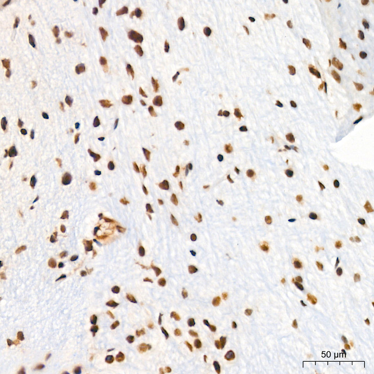
  • Immunohistochemistry analysis of ASH2L in paraffin-embedded mouse brain tissue using ASH2L Rabbit monoclonal antibody (STJ11102184) at a dilution of 1:200 (40x lens). High pressure antigen retrieval was performed with 0. 01 M citrate buffer (pH 6. 0) prior to immunohistochemistry staining.
  • Immunohistochemistry analysis of ASH2L in paraffin-embedded human colon carcinoma tissue using ASH2L Rabbit monoclonal antibody (STJ11102184) at a dilution of 1:200 (40x lens). High pressure antigen retrieval was performed with 0. 01 M citrate buffer (pH 6. 0) prior to immunohistochemistry staining.
  • Immunohistochemistry analysis of ASH2L in paraffin-embedded rat brain tissue using ASH2L Rabbit monoclonal antibody (STJ11102184) at a dilution of 1:200 (40x lens). High pressure antigen retrieval was performed with 0. 01 M citrate buffer (pH 6. 0) prior to immunohistochemistry staining.
  • Immunohistochemistry analysis of ASH2L in paraffin-embedded mouse lung tissue using ASH2L Rabbit monoclonal antibody (STJ11102184) at a dilution of 1:200 (40x lens). High pressure antigen retrieval was performed with 0. 01 M citrate buffer (pH 6. 0) prior to immunohistochemistry staining.
  • Western blot analysis of various lysates using ASH2L Rabbit monoclonal antibody (STJ11102184) at 1:1000 dilution. Secondary antibody: HRP Goat Anti-Rabbit IgG (H+L) (STJS000856) at 1:10000 dilution. Lysates/proteins: 25 Mu g per lane. Blocking buffer: 3% nonfat dry milk in TBST. Detection: ECL Basic Kit. Exposure time: 5s.

Anti-ASH2L antibody (529-628) [S4MR] (STJ11102184)

SKU:
STJ11102184

Shipping:
Free Shipping
Current Stock:
Host: Rabbit
Applications: WB/IHC/IF
Reactivity: Human/Mouse/Rat
Note: STRICTLY FOR FURTHER SCIENTIFIC RESEARCH USE ONLY (RUO). MUST NOT TO BE USED IN DIAGNOSTIC OR THERAPEUTIC APPLICATIONS.
Short Description: Rabbit monoclonal antibody anti-ASH2L (529-628) is suitable for use in Western Blot, Immunohistochemistry and Immunofluorescence research applications.
Clonality: Monoclonal
Clone ID: S4MR
Conjugation: Unconjugated
Isotype: IgG
Formulation: PBS with 0.02% Sodium Azide, 0.05% BSA, 50% Glycerol, pH7.3.
Purification: Affinity purification
Dilution Range: WB 1:500-1:1000
IHC-P 1:50-1:200
IF/ICC 1:50-1:200
Storage Instruction: Store at-20°C for up to 1 year from the date of receipt, and avoid repeat freeze-thaw cycles.
Gene Symbol: ASH2L
Gene ID: 9070
Uniprot ID: ASH2L_HUMAN
Immunogen Region: 529-628
Immunogen: A synthetic peptide corresponding to a sequence within amino acids 529-628 of human ASH2L (Q9UBL3).
Immunogen Sequence: AEKSLKQTPHSEIIFYKNGV NQGVAYKDIFEGVYFPAISL YKSCTVSINFGPCFKYPPKD LTYRPMSDMGWGAVVEHTLA DVLYHVETEVDGRRSPPWEP
Tissue Specificity Ubiquitously expressed. Predominantly expressed in adult heart and testis and fetal lung and liver, with barely detectable expression in adult lung, liver, kidney, prostate, and peripheral leukocytes.
Post Translational Modifications Both monomethylated and dimethylated on arginine residues in the C-terminus. Arg-296 is the major site. Methylation is not required for nuclear localization, nor for MLL complex integrity or maintenance of global histone H3K4me3 levels.
Function Transcriptional regulator. Component or associated component of some histone methyltransferase complexes which regulates transcription through recruitment of those complexes to gene promoters. Component of the Set1/Ash2 histone methyltransferase (HMT) complex, a complex that specifically methylates 'Lys-4' of histone H3, but not if the neighboring 'Lys-9' residue is already methylated. As part of the MLL1/MLL complex it is involved in methylation and dimethylation at 'Lys-4' of histone H3. May play a role in hematopoiesis. In association with RBBP5 and WDR5, stimulates the histone methyltransferase activities of KMT2A, KMT2B, KMT2C, KMT2D, SETD1A and SETD1B.
Protein Name Set1/Ash2 Histone Methyltransferase Complex Subunit Ash2
Ash2-Like Protein
Database Links Reactome: R-HSA-201722
Reactome: R-HSA-3214841
Reactome: R-HSA-3769402
Reactome: R-HSA-5617472
Reactome: R-HSA-8936459
Reactome: R-HSA-9772755
Cellular Localisation Nucleus
Alternative Antibody Names Anti-Set1/Ash2 Histone Methyltransferase Complex Subunit Ash2 antibody
Anti-Ash2-Like Protein antibody
Anti-ASH2L antibody
Anti-ASH2L1 antibody

Information sourced from Uniprot.org

12 months for antibodies. 6 months for ELISA Kits. Please see website T&Cs for further guidance