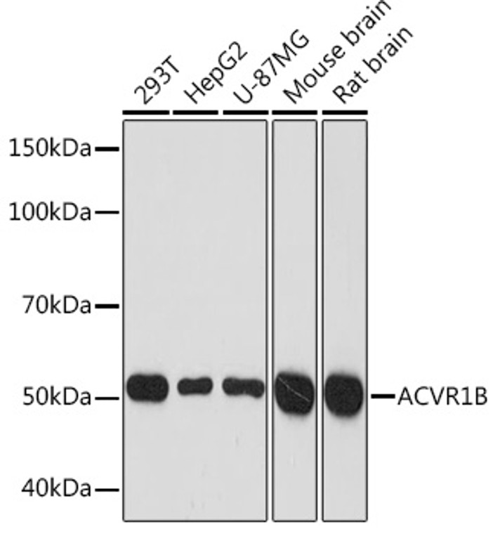
| Host: |
Rabbit |
| Applications: |
WB/IHC/IF |
| Reactivity: |
Human/Mouse/Rat |
| Note: |
STRICTLY FOR FURTHER SCIENTIFIC RESEARCH USE ONLY (RUO). MUST NOT TO BE USED IN DIAGNOSTIC OR THERAPEUTIC APPLICATIONS. |
| Short Description: |
Rabbit monoclonal antibody anti-ACVR1B (1-100) is suitable for use in Western Blot, Immunohistochemistry and Immunofluorescence research applications. |
| Clonality: |
Monoclonal |
| Clone ID: |
S4MR |
| Conjugation: |
Unconjugated |
| Isotype: |
IgG |
| Formulation: |
PBS with 0.02% Sodium Azide, 0.05% BSA, 50% Glycerol, pH7.3. |
| Purification: |
Affinity purification |
| Dilution Range: |
WB 1:500-1:2000IHC-P 1:50-1:200IF/ICC 1:50-1:200 |
| Storage Instruction: |
Store at-20°C for up to 1 year from the date of receipt, and avoid repeat freeze-thaw cycles. |
| Gene Symbol: |
ACVR1B |
| Gene ID: |
91 |
| Uniprot ID: |
ACV1B_HUMAN |
| Immunogen Region: |
1-100 |
| Immunogen: |
A synthetic peptide corresponding to a sequence within amino acids 1-100 of human ACVR1B (P36896). |
| Immunogen Sequence: |
MAESAGASSFFPLVVLLLAG SGGSGPRGVQALLCACTSCL QANYTCETDGACMVSIFNLD GMEHHVRTCIPKVELVPAGK PFYCLSSEDLRNTHCCYTDY |
| Tissue Specificity | Expressed in many tissues, most strongly in kidney, pancreas, brain, lung, and liver. |
| Post Translational Modifications | Autophosphorylated. Phosphorylated by activin receptor type-2 (ACVR2A or ACVR2B) in response to activin-binding at serine and threonine residues in the GS domain. Phosphorylation of ACVR1B by activin receptor type-2 regulates association with SMAD7. Ubiquitinated. Level of ubiquitination is regulated by the SMAD7-SMURF1 complex. Ubiquitinated. |
| Function | Transmembrane serine/threonine kinase activin type-1 receptor forming an activin receptor complex with activin receptor type-2 (ACVR2A or ACVR2B). Transduces the activin signal from the cell surface to the cytoplasm and is thus regulating a many physiological and pathological processes including neuronal differentiation and neuronal survival, hair follicle development and cycling, FSH production by the pituitary gland, wound healing, extracellular matrix production, immunosuppression and carcinogenesis. Activin is also thought to have a paracrine or autocrine role in follicular development in the ovary. Within the receptor complex, type-2 receptors (ACVR2A and/or ACVR2B) act as a primary activin receptors whereas the type-1 receptors like ACVR1B act as downstream transducers of activin signals. Activin binds to type-2 receptor at the plasma membrane and activates its serine-threonine kinase. The activated receptor type-2 then phosphorylates and activates the type-1 receptor such as ACVR1B. Once activated, the type-1 receptor binds and phosphorylates the SMAD proteins SMAD2 and SMAD3, on serine residues of the C-terminal tail. Soon after their association with the activin receptor and subsequent phosphorylation, SMAD2 and SMAD3 are released into the cytoplasm where they interact with the common partner SMAD4. This SMAD complex translocates into the nucleus where it mediates activin-induced transcription. Inhibitory SMAD7, which is recruited to ACVR1B through FKBP1A, can prevent the association of SMAD2 and SMAD3 with the activin receptor complex, thereby blocking the activin signal. Activin signal transduction is also antagonized by the binding to the receptor of inhibin-B via the IGSF1 inhibin coreceptor. ACVR1B also phosphorylates TDP2. |
| Protein Name | Activin Receptor Type-1bActivin Receptor Type IbActr-IbActivin Receptor-Like Kinase 4Alk-4Serine/Threonine-Protein Kinase Receptor R2Skr2 |
| Database Links | Reactome: R-HSA-1181150Reactome: R-HSA-1433617Reactome: R-HSA-1502540 |
| Cellular Localisation | Cell MembraneSingle-Pass Type I Membrane Protein |
| Alternative Antibody Names | Anti-Activin Receptor Type-1b antibodyAnti-Activin Receptor Type Ib antibodyAnti-Actr-Ib antibodyAnti-Activin Receptor-Like Kinase 4 antibodyAnti-Alk-4 antibodyAnti-Serine/Threonine-Protein Kinase Receptor R2 antibodyAnti-Skr2 antibodyAnti-ACVR1B antibodyAnti-ACVRLK4 antibodyAnti-ALK4 antibody |
Information sourced from Uniprot.org
12 months for antibodies. 6 months for ELISA Kits. Please see website T&Cs for further guidance


