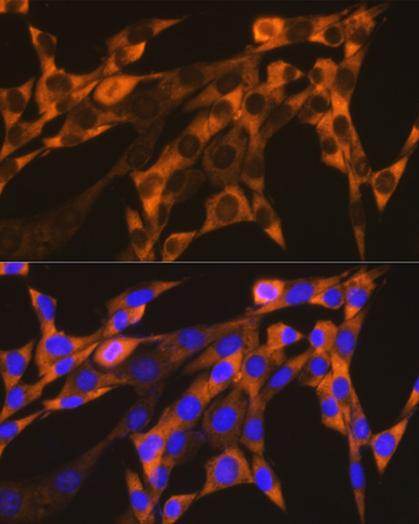
| Host: |
Rabbit |
| Applications: |
WB/IHC/IF |
| Reactivity: |
Human/Mouse/Rat |
| Note: |
STRICTLY FOR FURTHER SCIENTIFIC RESEARCH USE ONLY (RUO). MUST NOT TO BE USED IN DIAGNOSTIC OR THERAPEUTIC APPLICATIONS. |
| Short Description: |
Rabbit monoclonal antibody anti-YB1 (225-324) is suitable for use in Western Blot, Immunohistochemistry and Immunofluorescence research applications. |
| Clonality: |
Monoclonal |
| Clone ID: |
S3MR |
| Conjugation: |
Unconjugated |
| Isotype: |
IgG |
| Formulation: |
PBS with 0.02% Sodium Azide, 0.05% BSA, 50% Glycerol, pH7.3. |
| Purification: |
Affinity purification |
| Dilution Range: |
WB 1:500-1:2000IHC-P 1:50-1:200IF/ICC 1:50-1:200ChIP 1:500-1:1000 |
| Storage Instruction: |
Store at-20°C for up to 1 year from the date of receipt, and avoid repeat freeze-thaw cycles. |
| Gene Symbol: |
YBX1 |
| Gene ID: |
4904 |
| Uniprot ID: |
YBOX1_HUMAN |
| Immunogen Region: |
225-324 |
| Immunogen: |
A synthetic peptide corresponding to a sequence within amino acids 225-324 of human YB-1/YBX1 (P67809). |
| Immunogen Sequence: |
GAGEQGRPVRQNMYRGYRPR FRRGPPRQRQPREDGNEEDK ENQGDETQGQQPPQRRYRRN FNYRRRRPENPKPQDGKETK AADPPAENSSAPEAEQGGAE |
| Post Translational Modifications | Ubiquitinated by RBBP6.leading to a decrease of YBX1 transactivational ability. Phosphorylated.increased by TGFB1 treatment (Ref.6). Phosphorylation by PKB/AKT1 reduces interaction with cytoplasmic mRNA. In the absence of phosphorylation the protein is retained in the cytoplasm. Cleaved by a 20S proteasomal protease in response to agents that damage DNA. Cleavage takes place in the absence of ubiquitination and ATP. The resulting N-terminal fragment accumulates in the nucleus. |
| Function | DNA- and RNA-binding protein involved in various processes, such as translational repression, RNA stabilization, mRNA splicing, DNA repair and transcription regulation. Predominantly acts as a RNA-binding protein: binds preferentially to the 5'-CUCUGCG-3' RNA motif and specifically recognizes mRNA transcripts modified by C5-methylcytosine (m5C). Promotes mRNA stabilization: acts by binding to m5C-containing mRNAs and recruiting the mRNA stability maintainer ELAVL1, thereby preventing mRNA decay. Component of the CRD-mediated complex that promotes MYC mRNA stability. Contributes to the regulation of translation by modulating the interaction between the mRNA and eukaryotic initiation factors. Plays a key role in RNA composition of extracellular exosomes by defining the sorting of small non-coding RNAs, such as tRNAs, Y RNAs, Vault RNAs and miRNAs. Probably sorts RNAs in exosomes by recognizing and binding C5-methylcytosine (m5C)-containing RNAs. Acts as a key effector of epidermal progenitors by preventing epidermal progenitor senescence: acts by regulating the translation of a senescence-associated subset of cytokine mRNAs, possibly by binding to m5C-containing mRNAs. Also involved in pre-mRNA alternative splicing regulation: binds to splice sites in pre-mRNA and regulates splice site selection. Binds to TSC22D1 transcripts, thereby inhibiting their translation and negatively regulating TGF-beta-mediated transcription of COL1A2. Also able to bind DNA: regulates transcription of the multidrug resistance gene MDR1 is enhanced in presence of the APEX1 acetylated form at 'Lys-6' and 'Lys-7'. Binds to promoters that contain a Y-box (5'-CTGATTGGCCAA-3'), such as MDR1 and HLA class II genes. Promotes separation of DNA strands that contain mismatches or are modified by cisplatin. Has endonucleolytic activity and can introduce nicks or breaks into double-stranded DNA, suggesting a role in DNA repair. The secreted form acts as an extracellular mitogen and stimulates cell migration and proliferation. |
| Protein Name | Y-Box-Binding Protein 1Yb-1Ccaat-Binding Transcription Factor I Subunit ACbf-ADna-Binding Protein BDbpbEnhancer Factor I Subunit AEfi-ANuclease-Sensitive Element-Binding Protein 1Y-Box Transcription Factor |
| Database Links | Reactome: R-HSA-2173796Reactome: R-HSA-72163Reactome: R-HSA-72165Reactome: R-HSA-72203Reactome: R-HSA-877300Reactome: R-HSA-9017802 |
| Cellular Localisation | CytoplasmNucleusCytoplasmic GranuleSecretedExtracellular ExosomeP-BodyPredominantly Cytoplasmic In Proliferating CellsCytotoxic Stress And Dna Damage Enhance Translocation To The NucleusLocalized In Cytoplasmic Mrnp Granules Containing Untranslated MrnasShuttles Between Nucleus And CytoplasmLocalized With Ddx1Mbnl1 And Tial1 In Stress Granules Upon StressSecreted By Mesangial And Monocytic Cells After Inflammatory Challenges |
| Alternative Antibody Names | Anti-Y-Box-Binding Protein 1 antibodyAnti-Yb-1 antibodyAnti-Ccaat-Binding Transcription Factor I Subunit A antibodyAnti-Cbf-A antibodyAnti-Dna-Binding Protein B antibodyAnti-Dbpb antibodyAnti-Enhancer Factor I Subunit A antibodyAnti-Efi-A antibodyAnti-Nuclease-Sensitive Element-Binding Protein 1 antibodyAnti-Y-Box Transcription Factor antibodyAnti-YBX1 antibodyAnti-NSEP1 antibodyAnti-YB1 antibody |
Information sourced from Uniprot.org
12 months for antibodies. 6 months for ELISA Kits. Please see website T&Cs for further guidance


![Confocal imaging of HeLa cells using [KO Validated] CDKN2A/p16INK4a Rabbit monoclonal antibody (STJ113253, dilution 1:200) followed by a further incubation with Cy3 Goat Anti-Rabbit IgG (H+L) (dilution 1:500) (Red). The cells were counterstained with Alpha-Tubulin Mouse monoclonal antibody ( dilution 1:400) followed by incubation with ABflo® 488-conjugated Goat Anti-Mouse IgG (H+L) antibody ( dilution 1:500) (Green). DAPI was used for nuclear staining (Blue). Objective: 100x. Confocal imaging of HeLa cells using [KO Validated] CDKN2A/p16INK4a Rabbit monoclonal antibody (STJ113253, dilution 1:200) followed by a further incubation with Cy3 Goat Anti-Rabbit IgG (H+L) (dilution 1:500) (Red). The cells were counterstained with Alpha-Tubulin Mouse monoclonal antibody ( dilution 1:400) followed by incubation with ABflo® 488-conjugated Goat Anti-Mouse IgG (H+L) antibody ( dilution 1:500) (Green). DAPI was used for nuclear staining (Blue). Objective: 100x.](https://cdn11.bigcommerce.com/s-zso2xnchw9/images/stencil/300x300/products/92819/373410/STJ113253_1__34054.1713139652.jpg?c=1)