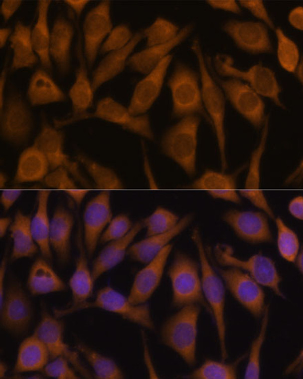
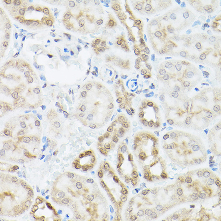
| Host: |
Rabbit |
| Applications: |
WB/IHC/IF |
| Reactivity: |
Human/Mouse |
| Note: |
STRICTLY FOR FURTHER SCIENTIFIC RESEARCH USE ONLY (RUO). MUST NOT TO BE USED IN DIAGNOSTIC OR THERAPEUTIC APPLICATIONS. |
| Short Description: |
Rabbit polyclonal antibody anti-UBQLN2 (1-150) is suitable for use in Western Blot, Immunohistochemistry and Immunofluorescence research applications. |
| Clonality: |
Polyclonal |
| Conjugation: |
Unconjugated |
| Isotype: |
IgG |
| Formulation: |
PBS with 0.02% Sodium Azide, 50% Glycerol, pH7.3. |
| Purification: |
Affinity purification |
| Dilution Range: |
WB 1:200-1:2000IHC-P 1:50-1:200IF/ICC 1:50-1:200 |
| Storage Instruction: |
Store at-20°C for up to 1 year from the date of receipt, and avoid repeat freeze-thaw cycles. |
| Gene Symbol: |
UBQLN2 |
| Gene ID: |
29978 |
| Uniprot ID: |
UBQL2_HUMAN |
| Immunogen Region: |
1-150 |
| Immunogen: |
Recombinant fusion protein containing a sequence corresponding to amino acids 1-150 of human UBQLN2 (NP_038472.2). |
| Immunogen Sequence: |
MAENGESSGPPRPSRGPAAA QGSAAAPAEPKIIKVTVKTP KEKEEFAVPENSSVQQFKEA ISKRFKSQTDQLVLIFAGKI LKDQDTLIQHGIHDGLTVHL VIKSQNRPQGQSTQPSNAAG TNTTSASTPRSNSTPISTNS NPFGLGSLGG |
| Post Translational Modifications | Degraded during macroautophagy. |
| Function | Plays an important role in the regulation of different protein degradation mechanisms and pathways including ubiquitin-proteasome system (UPS), autophagy and the endoplasmic reticulum-associated protein degradation (ERAD) pathway. Mediates the proteasomal targeting of misfolded or accumulated proteins for degradation by binding (via UBA domain) to their polyubiquitin chains and by interacting (via ubiquitin-like domain) with the subunits of the proteasome. Plays a role in the ERAD pathway via its interaction with ER-localized proteins FAF2/UBXD8 and HERPUD1 and may form a link between the polyubiquitinated ERAD substrates and the proteasome. Involved in the regulation of macroautophagy and autophagosome formation.required for maturation of autophagy-related protein LC3 from the cytosolic form LC3-I to the membrane-bound form LC3-II and may assist in the maturation of autophagosomes to autolysosomes by mediating autophagosome-lysosome fusion. Negatively regulates the endocytosis of GPCR receptors: AVPR2 and ADRB2, by specifically reducing the rate at which receptor-arrestin complexes concentrate in clathrin-coated pits (CCPs). |
| Protein Name | Ubiquilin-2Chap1Dsk2 HomologProtein Linking Iap With Cytoskeleton 2Plic-2Hplic-2Ubiquitin-Like Product Chap1/Dsk2 |
| Database Links | Reactome: R-HSA-8856825 |
| Cellular Localisation | CytoplasmNucleusMembraneCytoplasmic VesicleAutophagosomeColocalizes With A Subset Of ProteasomesNamely Those That Are Cytoskeleton Associated Or Free In The CytosolAssociated With Fibers In Mitotic Cells |
| Alternative Antibody Names | Anti-Ubiquilin-2 antibodyAnti-Chap1 antibodyAnti-Dsk2 Homolog antibodyAnti-Protein Linking Iap With Cytoskeleton 2 antibodyAnti-Plic-2 antibodyAnti-Hplic-2 antibodyAnti-Ubiquitin-Like Product Chap1/Dsk2 antibodyAnti-UBQLN2 antibodyAnti-N4BP4 antibodyAnti-PLIC2 antibodyAnti-HRIHFB2157 antibody |
Information sourced from Uniprot.org
12 months for antibodies. 6 months for ELISA Kits. Please see website T&Cs for further guidance


