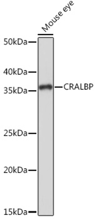
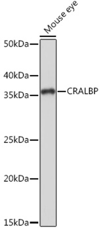
• Western blot analysis of extracts of mouse eye, using CRALBP rabbit monoclonal antibody (STJ11102467) at 1:1000 dilution. Secondary antibody: HRP Goat Anti-rabbit IgG (H+L) at 1:10000 dilution. Lysates/proteins: 25ug per lane. Blocking buffer: 3% non-fat dry milk in TBST. Detection: ECL Basic Kit. Exposure time: 1s.
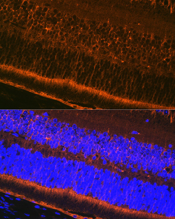
  • Immunofluorescence analysis of paraffin-embedded mouse eye using CRALBP Rabbit monoclonal antibody (STJ11102467) at dilution of 1:100 (40x lens). Secondary antibody: Cy3 Goat Anti-Rabbit IgG (H+L) at 1:500 dilution. Blue: DAPI for nuclear staining.
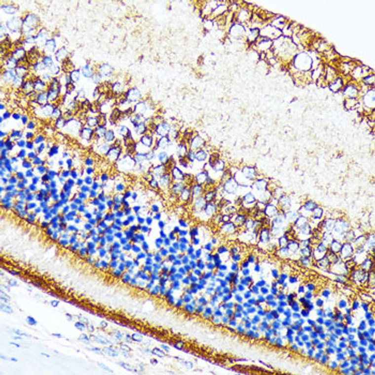
  • Immunohistochemistry of paraffin-embedded rat retina using CRALBP rabbit monoclonal antibody (STJ11102467) at dilution of 1:100 (40x lens). Perform microwave antigen retrieval with 10 mM Tris/EDTA buffer pH 9. 0 before commencing with immunohistochemistry staining protocol.
  • Immunofluorescence analysis of paraffin-embedded rat eye using CRALBP Rabbit monoclonal antibody (STJ11102467) at dilution of 1:100 (40x lens). Secondary antibody: Cy3 Goat Anti-Rabbit IgG (H+L) at 1:500 dilution. Blue: DAPI for nuclear staining.
  • Immunohistochemistry of paraffin-embedded mouse retina using CRALBP rabbit monoclonal antibody (STJ11102467) at dilution of 1:100 (40x lens). Perform microwave antigen retrieval with 10 mM Tris/EDTA buffer pH 9. 0 before commencing with immunohistochemistry staining protocol.
  • Immunohistochemistry analysis of CRALBP in paraffin-embedded mouse retina using CRALBP Rabbit monoclonal antibody (STJ11102467) at dilution of 1:100 (40x lens). Perform microwave antigen retrieval with 10 mM Tris/EDTA buffer pH 9. 0 before commencing with immunohistochemistry staining protocol.
  • Immunofluorescence analysis of rat eye using CRALBP rabbit monoclonal antibody (STJ11102467) at dilution of 1:100 (40x lens). Blue: DAPI for nuclear staining.
  • Immunohistochemistry analysis of CRALBP in paraffin-embedded rat retina using CRALBP Rabbit monoclonal antibody (STJ11102467) at dilution of 1:100 (40x lens). Perform microwave antigen retrieval with 10 mM Tris/EDTA buffer pH 9. 0 before commencing with immunohistochemistry staining protocol.
  • Immunofluorescence analysis of mouse eye using CRALBP rabbit monoclonal antibody (STJ11102467) at dilution of 1:100 (40x lens). Blue: DAPI for nuclear staining.
  • Western blot analysis of lysates from Mouse eye, using CRALBP Rabbit monoclonal antibody (STJ11102467) at 1:1000 dilution. Secondary antibody: HRP Goat Anti-Rabbit IgG (H+L) (STJS000856) at 1:10000 dilution. Lysates/proteins: 25 Mu g per lane. Blocking buffer: 3% nonfat dry milk in TBST. Detection: ECL Basic Kit. Exposure time: 1s.

Anti-RLBP1 antibody (1-100) [S7MR] (STJ11102467)

SKU:
STJ11102467

Shipping:
Free Shipping
Current Stock:
Host: Rabbit
Applications: WB/IHC/IF
Reactivity: Human/Mouse/Rat
Note: STRICTLY FOR FURTHER SCIENTIFIC RESEARCH USE ONLY (RUO). MUST NOT TO BE USED IN DIAGNOSTIC OR THERAPEUTIC APPLICATIONS.
Short Description: Rabbit monoclonal antibody anti-CRALBP (1-100) is suitable for use in Western Blot, Immunohistochemistry and Immunofluorescence research applications.
Clonality: Monoclonal
Clone ID: S7MR
Conjugation: Unconjugated
Isotype: IgG
Formulation: PBS with 0.02% Sodium Azide, 0.05% BSA, 50% Glycerol, pH7.3.
Purification: Affinity purification
Dilution Range: WB 1:500-1:2000
IHC-P 1:50-1:200
IF/ICC 1:50-1:200
Storage Instruction: Store at-20°C for up to 1 year from the date of receipt, and avoid repeat freeze-thaw cycles.
Gene Symbol: RLBP1
Gene ID: 6017
Uniprot ID: RLBP1_HUMAN
Immunogen Region: 1-100
Immunogen: A synthetic peptide corresponding to a sequence within amino acids 1-100 of human CRALBP (P12271).
Immunogen Sequence: MSEGVGTFRMVPEEEQELRA QLEQLTTKDHGPVFGPCSQL PRHTLQKAKDELNEREETRE EAVRELQEMVQAQAASGEEL AVAVAERVQEKDSGFFLRFI
Tissue Specificity Retina and pineal gland. Not present in photoreceptor cells but is expressed abundantly in the adjacent retinal pigment epithelium (RPE) and in the Mueller glial cells of the retina.
Function Soluble retinoid carrier essential the proper function of both rod and cone photoreceptors. Participates in the regeneration of active 11-cis-retinol and 11-cis-retinaldehyde, from the inactive 11-trans products of the rhodopsin photocycle and in the de novo synthesis of these retinoids from 11-trans metabolic precursors. The cycling of retinoids between photoreceptor and adjacent pigment epithelium cells is known as the 'visual cycle'.
Protein Name Retinaldehyde-Binding Protein 1
Cellular Retinaldehyde-Binding Protein
Database Links Reactome: R-HSA-2187335
Reactome: R-HSA-2453864
Reactome: R-HSA-2453902
Cellular Localisation Cytoplasm
Alternative Antibody Names Anti-Retinaldehyde-Binding Protein 1 antibody
Anti-Cellular Retinaldehyde-Binding Protein antibody
Anti-RLBP1 antibody
Anti-CRALBP antibody

Information sourced from Uniprot.org

12 months for antibodies. 6 months for ELISA Kits. Please see website T&Cs for further guidance