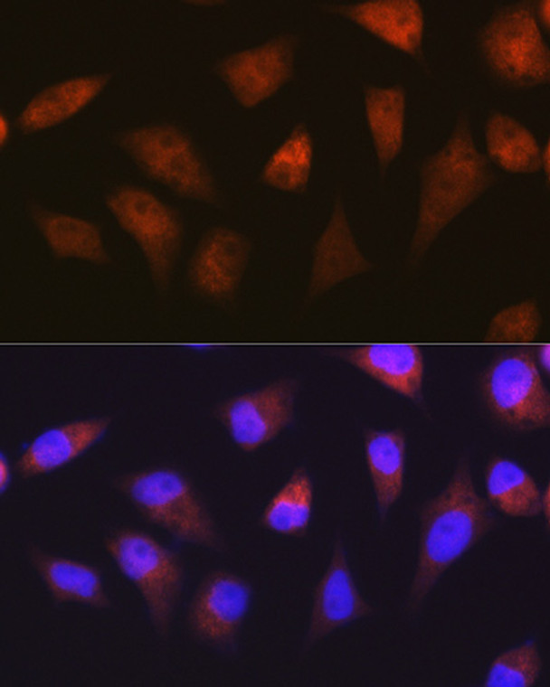
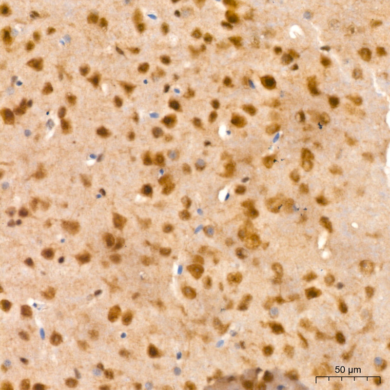
| Host: | Rabbit |
| Applications: | WB/IHC/IF |
| Reactivity: | Human/Mouse/Rat |
| Note: | STRICTLY FOR FURTHER SCIENTIFIC RESEARCH USE ONLY (RUO). MUST NOT TO BE USED IN DIAGNOSTIC OR THERAPEUTIC APPLICATIONS. |
| Short Description: | Rabbit monoclonal antibody anti-PSMA2 (1-100) is suitable for use in Western Blot, Immunohistochemistry and Immunofluorescence research applications. |
| Clonality: | Monoclonal |
| Clone ID: | S7MR |
| Conjugation: | Unconjugated |
| Isotype: | IgG |
| Formulation: | PBS with 0.02% Sodium Azide, 0.05% BSA, 50% Glycerol, pH7.3. |
| Purification: | Affinity purification |
| Dilution Range: | WB 1:500-1:2000IHC-P 1:50-1:200IF/ICC 1:50-1:200 |
| Storage Instruction: | Store at-20°C for up to 1 year from the date of receipt, and avoid repeat freeze-thaw cycles. |
| Gene Symbol: | PSMA2 |
| Gene ID: | 5683 |
| Uniprot ID: | PSA2_HUMAN |
| Immunogen Region: | 1-100 |
| Immunogen: | Recombinant fusion protein containing a sequence corresponding to amino acids 1-100 of human PSMA2 (P25787). |
| Immunogen Sequence: | MAERGYSFSLTTFSPSGKLV QIEYALAAVAGGAPSVGIKA ANGVVLATEKKQKSILYDER SVHKVEPITKHIGLVYSGMG PDYRVLVHRARKLAQQYYLV |
| Post Translational Modifications | Phosphorylated on tyrosine residues.which may be important for nuclear import. |
| Function | Component of the 20S core proteasome complex involved in the proteolytic degradation of most intracellular proteins. This complex plays numerous essential roles within the cell by associating with different regulatory particles. Associated with two 19S regulatory particles, forms the 26S proteasome and thus participates in the ATP-dependent degradation of ubiquitinated proteins. The 26S proteasome plays a key role in the maintenance of protein homeostasis by removing misfolded or damaged proteins that could impair cellular functions, and by removing proteins whose functions are no longer required. Associated with the PA200 or PA28, the 20S proteasome mediates ubiquitin-independent protein degradation. This type of proteolysis is required in several pathways including spermatogenesis (20S-PA200 complex) or generation of a subset of MHC class I-presented antigenic peptides (20S-PA28 complex). |
| Protein Name | Proteasome Subunit Alpha Type-2Macropain Subunit C3Multicatalytic Endopeptidase Complex Subunit C3Proteasome Component C3 |
| Database Links | Reactome: R-HSA-1169091Reactome: R-HSA-1234176Reactome: R-HSA-1236974Reactome: R-HSA-1236978Reactome: R-HSA-174084Reactome: R-HSA-174113Reactome: R-HSA-174154Reactome: R-HSA-174178Reactome: R-HSA-174184Reactome: R-HSA-180534Reactome: R-HSA-180585Reactome: R-HSA-187577Reactome: R-HSA-195253Reactome: R-HSA-202424Reactome: R-HSA-211733Reactome: R-HSA-2467813Reactome: R-HSA-2871837Reactome: R-HSA-349425Reactome: R-HSA-350562Reactome: R-HSA-382556Reactome: R-HSA-450408Reactome: R-HSA-4608870Reactome: R-HSA-4641257Reactome: R-HSA-4641258Reactome: R-HSA-5358346Reactome: R-HSA-5362768Reactome: R-HSA-5607761Reactome: R-HSA-5607764Reactome: R-HSA-5610780Reactome: R-HSA-5610783Reactome: R-HSA-5610785Reactome: R-HSA-5632684Reactome: R-HSA-5658442Reactome: R-HSA-5668541Reactome: R-HSA-5676590Reactome: R-HSA-5678895Reactome: R-HSA-5687128Reactome: R-HSA-5689603Reactome: R-HSA-5689880Reactome: R-HSA-6798695Reactome: R-HSA-68867Reactome: R-HSA-68949Reactome: R-HSA-69017Reactome: R-HSA-69481Reactome: R-HSA-69601Reactome: R-HSA-75815Reactome: R-HSA-8852276Reactome: R-HSA-8854050Reactome: R-HSA-8939236Reactome: R-HSA-8939902Reactome: R-HSA-8941858Reactome: R-HSA-8948751Reactome: R-HSA-8951664Reactome: R-HSA-9010553Reactome: R-HSA-9020702Reactome: R-HSA-9604323Reactome: R-HSA-9755511Reactome: R-HSA-9762114Reactome: R-HSA-9824272Reactome: R-HSA-983168 |
| Cellular Localisation | CytoplasmNucleusTranslocated From The Cytoplasm Into The Nucleus Following Interaction With Akirin2Which Bridges The Proteasome With The Nuclear Import Receptor Ipo9Colocalizes With Trim5 In Cytoplasmic Bodies |
| Alternative Antibody Names | Anti-Proteasome Subunit Alpha Type-2 antibodyAnti-Macropain Subunit C3 antibodyAnti-Multicatalytic Endopeptidase Complex Subunit C3 antibodyAnti-Proteasome Component C3 antibodyAnti-PSMA2 antibodyAnti-HC3 antibodyAnti-PSC3 antibody |
Information sourced from Uniprot.org
12 months for antibodies. 6 months for ELISA Kits. Please see website T&Cs for further guidance


