| Host: |
Rabbit |
| Applications: |
WB |
| Reactivity: |
Mouse/Rat |
| Note: |
STRICTLY FOR FURTHER SCIENTIFIC RESEARCH USE ONLY (RUO). MUST NOT TO BE USED IN DIAGNOSTIC OR THERAPEUTIC APPLICATIONS. |
| Short Description: |
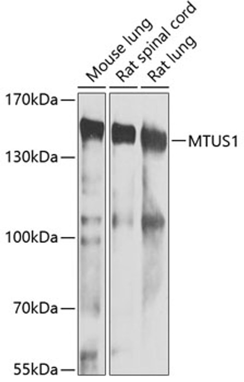
Rabbit polyclonal antibody anti-MTUS1 (207-436) is suitable for use in Western Blot research applications. |
| Clonality: |
Polyclonal |
| Conjugation: |
Unconjugated |
| Isotype: |
IgG |
| Formulation: |
PBS with 0.02% Sodium Azide, 50% Glycerol, pH7.3. |
| Purification: |
Affinity purification |
| Dilution Range: |
WB 1:500-1:2000 |
| Storage Instruction: |
Store at-20°C for up to 1 year from the date of receipt, and avoid repeat freeze-thaw cycles. |
| Gene Symbol: |
MTUS1 |
| Gene ID: |
57509 |
| Uniprot ID: |
MTUS1_HUMAN |
| Immunogen Region: |
207-436 |
| Immunogen: |
Recombinant fusion protein containing a sequence corresponding to amino acids 207-436 of human MTUS1 (NP_065800.1). |
| Immunogen Sequence: |
NAAHETSKLEIEASHSEKLE LLKKAYEASLSEIKKGHEIE KKSLEDLLSEKQESLEKQIN DLKSENDALNEKLKSEEQKR RAREKANLKNPQIMYLEQEL ESLKAVLEIKNEKLHQQDIK LMKMEKLVDNNTALVDKLKR FQQENEELKARMDKHMAISR QLSTEQAVLQESLEKESKVN KRLSMENEELLWKLHNGDLC SPKRSPTSSAIPLQSPRNSG SFPSPSISPR |
| Tissue Specificity | Ubiquitously expressed (at protein level). Highly expressed in brain. Down-regulated in ovarian carcinoma, pancreas carcinoma, colon carcinoma and head and neck squamous cell carcinoma (HNSCC). Isoform 1 is the major isoform in most peripheral tissues. Isoform 2 is abundant in most peripheral tissues. Isoform 3 is the major isoform in brain, female reproductive tissues, thyroid and heart. Within brain it is highly expressed in corpus callosum and pons. Isoform 6 is brain-specific, it is the major isoform in cerebellum and fetal brain. |
| Function | Cooperates with AGTR2 to inhibit ERK2 activation and cell proliferation. May be required for AGTR2 cell surface expression. Together with PTPN6, induces UBE2V2 expression upon angiotensin-II stimulation. Isoform 1 inhibits breast cancer cell proliferation, delays the progression of mitosis by prolonging metaphase and reduces tumor growth. |
| Protein Name | Microtubule-Associated Tumor Suppressor 1At2 Receptor-Binding ProteinAngiotensin-Ii Type 2 Receptor-Interacting ProteinMitochondrial Tumor Suppressor 1 |
| Cellular Localisation | MitochondrionGolgi ApparatusCell MembraneNucleusIn NeuronsTranslocates Into The Nucleus After Treatment With Angiotensin-IiIsoform 1: CytoplasmCytoskeletonMicrotubule Organizing CenterCentrosomeCytoplasmSpindleLocalizes With The Mitotic Spindle During Mitosis And With The Intercellular Bridge During Cytokinesis |
| Alternative Antibody Names | Anti-Microtubule-Associated Tumor Suppressor 1 antibodyAnti-At2 Receptor-Binding Protein antibodyAnti-Angiotensin-Ii Type 2 Receptor-Interacting Protein antibodyAnti-Mitochondrial Tumor Suppressor 1 antibodyAnti-MTUS1 antibodyAnti-ATBP antibodyAnti-ATIP antibodyAnti-GK1 antibodyAnti-KIAA1288 antibodyAnti-MTSG1 antibody |
Information sourced from Uniprot.org
12 months for antibodies. 6 months for ELISA Kits. Please see website T&Cs for further guidance


