| Host: |
Rabbit |
| Applications: |
WB/FC/IHC |
| Reactivity: |
Human/Mouse/Rat |
| Note: |
STRICTLY FOR FURTHER SCIENTIFIC RESEARCH USE ONLY (RUO). MUST NOT TO BE USED IN DIAGNOSTIC OR THERAPEUTIC APPLICATIONS. |
| Short Description: |
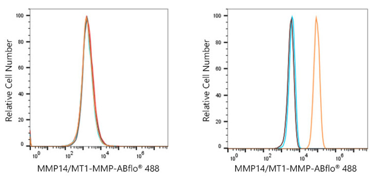
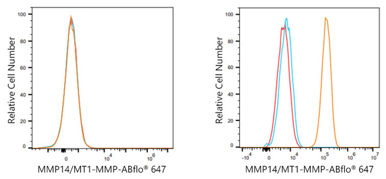
Rabbit monoclonal antibody anti-MT1-MMP (100-200) is suitable for use in Western Blot, Flow Cytometry and Immunohistochemistry research applications. |
| Clonality: |
Monoclonal |
| Clone ID: |
S4MR |
| Conjugation: |
Unconjugated |
| Isotype: |
IgG |
| Formulation: |
PBS with 0.02% Sodium Azide, 0.05% BSA, 50% Glycerol, pH7.3. |
| Purification: |
Affinity purification |
| Dilution Range: |
WB 1:500-1:2000FC 1:100-1:500IHC-P 1:50-1:200 |
| Storage Instruction: |
Store at-20°C for up to 1 year from the date of receipt, and avoid repeat freeze-thaw cycles. |
| Gene Symbol: |
MMP14 |
| Gene ID: |
4323 |
| Uniprot ID: |
MMP14_HUMAN |
| Immunogen Region: |
100-200 |
| Immunogen: |
A synthetic peptide corresponding to a sequence within amino acids 100-200 of human MMP14/MMP14/MT1-MMP (P50281). |
| Immunogen Sequence: |
GAEIKANVRRKRYAIQGLKW QHNEITFCIQNYTPKVGEYA TYEAIRKAFRVWESATPLRF REVPYAYIREGHEKQADIMI FFAEGFHGDSTPFDGEGGFL A |
| Tissue Specificity | Expressed in stromal cells of colon, breast, and head and neck. Expressed in lung tumors. |
| Post Translational Modifications | The precursor is cleaved by a furin endopeptidase. Tyrosine phosphorylated by PKDCC/VLK. |
| Function | Endopeptidase that degrades various components of the extracellular matrix such as collagen. Activates progelatinase A. Essential for pericellular collagenolysis and modeling of skeletal and extraskeletal connective tissues during development. May be involved in actin cytoskeleton reorganization by cleaving PTK7. Acts as a positive regulator of cell growth and migration via activation of MMP15. Involved in the formation of the fibrovascular tissues in association with pro-MMP2. Cleaves ADGRB1 to release vasculostatin-40 which inhibits angiogenesis. |
| Protein Name | Matrix Metalloproteinase-14Mmp-14Mmp-X1Membrane-Type Matrix Metalloproteinase 1Mt-Mmp 1Mtmmp1Membrane-Type-1 Matrix MetalloproteinaseMt1-MmpMt1mmp |
| Database Links | Reactome: R-HSA-1442490Reactome: R-HSA-1474228Reactome: R-HSA-1592389 |
| Cellular Localisation | MembraneSingle-Pass Type I Membrane ProteinMelanosomeCytoplasmIdentified By Mass Spectrometry In Melanosome Fractions From Stage I To Stage IvForms A Complex With Bst2 And Localizes To The Cytoplasm |
| Alternative Antibody Names | Anti-Matrix Metalloproteinase-14 antibodyAnti-Mmp-14 antibodyAnti-Mmp-X1 antibodyAnti-Membrane-Type Matrix Metalloproteinase 1 antibodyAnti-Mt-Mmp 1 antibodyAnti-Mtmmp1 antibodyAnti-Membrane-Type-1 Matrix Metalloproteinase antibodyAnti-Mt1-Mmp antibodyAnti-Mt1mmp antibodyAnti-MMP14 antibody |
Information sourced from Uniprot.org
12 months for antibodies. 6 months for ELISA Kits. Please see website T&Cs for further guidance


