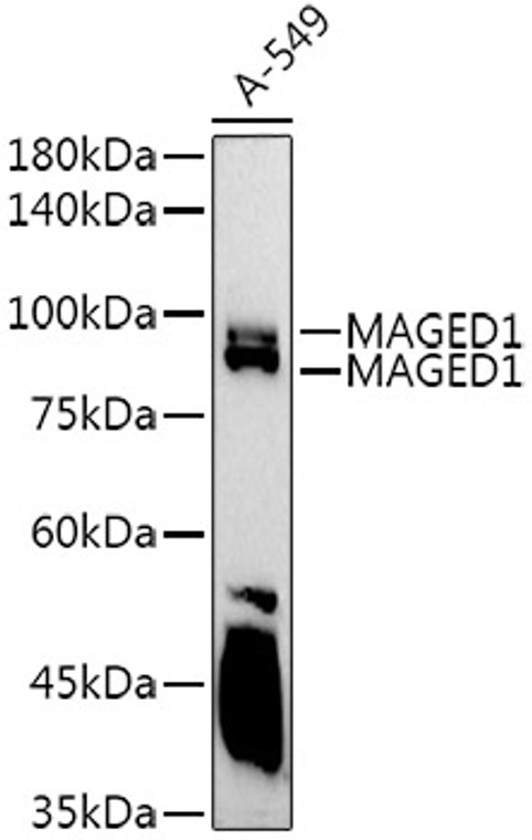
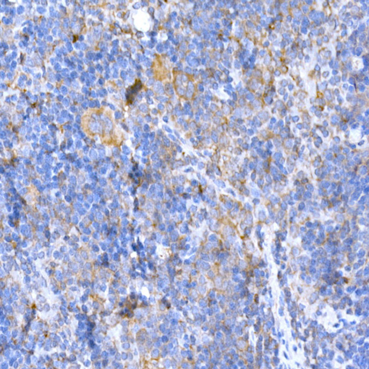
| Host: |
Rabbit |
| Applications: |
WB/IHC |
| Reactivity: |
Human/Mouse/Rat |
| Note: |
STRICTLY FOR FURTHER SCIENTIFIC RESEARCH USE ONLY (RUO). MUST NOT TO BE USED IN DIAGNOSTIC OR THERAPEUTIC APPLICATIONS. |
| Short Description: |
Rabbit polyclonal antibody anti-MAGED1 (635-834) is suitable for use in Western Blot and Immunohistochemistry research applications. |
| Clonality: |
Polyclonal |
| Conjugation: |
Unconjugated |
| Isotype: |
IgG |
| Formulation: |
PBS with 0.02% Sodium Azide, 50% Glycerol, pH7.3. |
| Purification: |
Affinity purification |
| Dilution Range: |
WB 1:100-1:500IHC-P 1:100-1:500 |
| Storage Instruction: |
Store at-20°C for up to 1 year from the date of receipt, and avoid repeat freeze-thaw cycles. |
| Gene Symbol: |
MAGED1 |
| Gene ID: |
9500 |
| Uniprot ID: |
MAGD1_HUMAN |
| Immunogen Region: |
635-834 |
| Immunogen: |
Recombinant fusion protein containing a sequence corresponding to amino acids 635-834 of human MAGED1/NRAGE (NP_001005333.1). |
| Immunogen Sequence: |
EAVLWEALRKMGLRPGVRHP LLGDLRKLLTYEFVKQKYLD YRRVPNSNPPEYEFLWGLRS YHETSKMKVLRFIAEVQKRD PRDWTAQFMEAADEALDALD AAAAEAEARAEARTRMGIGD EAVSGPWSWDDIEFELLTWD EEGDFGDPWSRIPFTFWARY HQNARSRFPQTFAGPIIGPG GTASANFAANFGAIGFFWVE |
| Tissue Specificity | Expressed in bone marrow stromal cells from both multiple myeloma patients and healthy donors. Seems to be ubiquitously expressed. |
| Function | Involved in the apoptotic response after nerve growth factor (NGF) binding in neuronal cells. Inhibits cell cycle progression, and facilitates NGFR-mediated apoptosis. May act as a regulator of the function of DLX family members. May enhance ubiquitin ligase activity of RING-type zinc finger-containing E3 ubiquitin-protein ligases. Proposed to act through recruitment and/or stabilization of the Ubl-conjugating enzyme (E2) at the E3:substrate complex. Plays a role in the circadian rhythm regulation. May act as RORA co-regulator, modulating the expression of core clock genes such as BMAL1 and NFIL3, induced, or NR1D1, repressed. |
| Protein Name | Melanoma-Associated Antigen D1Mage Tumor Antigen CcfMage-D1 AntigenNeurotrophin Receptor-Interacting Mage Homolog |
| Database Links | Reactome: R-HSA-193648Reactome: R-HSA-418889Reactome: R-HSA-9768919 |
| Cellular Localisation | CytoplasmCell MembranePeripheral Membrane ProteinNucleusExpression Shifts From The Cytoplasm To The Plasma Membrane Upon Stimulation With Ngf |
| Alternative Antibody Names | Anti-Melanoma-Associated Antigen D1 antibodyAnti-Mage Tumor Antigen Ccf antibodyAnti-Mage-D1 Antigen antibodyAnti-Neurotrophin Receptor-Interacting Mage Homolog antibodyAnti-MAGED1 antibodyAnti-NRAGE antibodyAnti-PP2250 antibodyAnti-PRO2292 antibody |
Information sourced from Uniprot.org
12 months for antibodies. 6 months for ELISA Kits. Please see website T&Cs for further guidance


