| Host: |
Rabbit |
| Applications: |
WB |
| Reactivity: |
Human/Mouse |
| Note: |
STRICTLY FOR FURTHER SCIENTIFIC RESEARCH USE ONLY (RUO). MUST NOT TO BE USED IN DIAGNOSTIC OR THERAPEUTIC APPLICATIONS. |
| Short Description: |
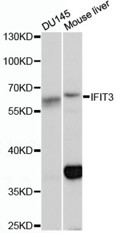
Rabbit polyclonal antibody anti-IFIT3 (291-490) is suitable for use in Western Blot research applications. |
| Clonality: |
Polyclonal |
| Conjugation: |
Unconjugated |
| Isotype: |
IgG |
| Formulation: |
PBS with 0.02% Sodium Azide, 50% Glycerol, pH7.3. |
| Purification: |
Affinity purification |
| Dilution Range: |
WB 1:500-1:2000 |
| Storage Instruction: |
Store at-20°C for up to 1 year from the date of receipt, and avoid repeat freeze-thaw cycles. |
| Gene Symbol: |
IFIT3 |
| Gene ID: |
3437 |
| Uniprot ID: |
IFIT3_HUMAN |
| Immunogen Region: |
291-490 |
| Immunogen: |
Recombinant fusion protein containing a sequence corresponding to amino acids 291-490 of human IFIT3 (NP_001540.2). |
| Immunogen Sequence: |
QMQNTGESEASGNKEMIEAL KQYAMDYSNKALEKGLNPLN AYSDLAEFLETECYQTPFNK EVPDAEKQQSHQRYCNLQKY NGKSEDTAVQHGLEGLSISK KSTDKEEIKDQPQNVSENLL PQNAPNYWYLQGLIHKQNGD LLQAAKCYEKELGRLLRDAP SGIGSIFLSASELEDGSEEM GQGAVSSSPRELLSNSEQLN |
| Tissue Specificity | Expression significantly higher in peripheral blood mononuclear cells (PBMCs) and monocytes from systemic lupus erythematosus (SLE) patients than in those from healthy individuals (at protein level). Spleen, lung, leukocytes, lymph nodes, placenta, bone marrow and fetal liver. |
| Function | IFN-induced antiviral protein which acts as an inhibitor of cellular as well as viral processes, cell migration, proliferation, signaling, and viral replication. Enhances MAVS-mediated host antiviral responses by serving as an adapter bridging TBK1 to MAVS which leads to the activation of TBK1 and phosphorylation of IRF3 and phosphorylated IRF3 translocates into nucleus to promote antiviral gene transcription. Exhibits an antiproliferative activity via the up-regulation of cell cycle negative regulators CDKN1A/p21 and CDKN1B/p27. Normally, CDKN1B/p27 turnover is regulated by COPS5, which binds CDKN1B/p27 in the nucleus and exports it to the cytoplasm for ubiquitin-dependent degradation. IFIT3 sequesters COPS5 in the cytoplasm, thereby increasing nuclear CDKN1B/p27 protein levels. Up-regulates CDKN1A/p21 by down-regulating MYC, a repressor of CDKN1A/p21. Can negatively regulate the apoptotic effects of IFIT2. |
| Protein Name | Interferon-Induced Protein With Tetratricopeptide Repeats 3Ifit-3Cig49Isg-60Interferon-Induced 60 Kda ProteinIfi-60kInterferon-Induced Protein With Tetratricopeptide Repeats 4Ifit-4Retinoic Acid-Induced Gene G ProteinP60Rig-G |
| Database Links | Reactome: R-HSA-909733 |
| Cellular Localisation | CytoplasmMitochondrion |
| Alternative Antibody Names | Anti-Interferon-Induced Protein With Tetratricopeptide Repeats 3 antibodyAnti-Ifit-3 antibodyAnti-Cig49 antibodyAnti-Isg-60 antibodyAnti-Interferon-Induced 60 Kda Protein antibodyAnti-Ifi-60k antibodyAnti-Interferon-Induced Protein With Tetratricopeptide Repeats 4 antibodyAnti-Ifit-4 antibodyAnti-Retinoic Acid-Induced Gene G Protein antibodyAnti-P60 antibodyAnti-Rig-G antibodyAnti-IFIT3 antibodyAnti-CIG-49 antibodyAnti-IFI60 antibodyAnti-IFIT4 antibodyAnti-ISG60 antibody |
Information sourced from Uniprot.org
12 months for antibodies. 6 months for ELISA Kits. Please see website T&Cs for further guidance


