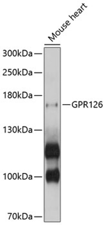
| Host: |
Rabbit |
| Applications: |
WB |
| Reactivity: |
Mouse |
| Note: |
STRICTLY FOR FURTHER SCIENTIFIC RESEARCH USE ONLY (RUO). MUST NOT TO BE USED IN DIAGNOSTIC OR THERAPEUTIC APPLICATIONS. |
| Short Description: |
Rabbit polyclonal antibody anti-GPR126 (40-300) is suitable for use in Western Blot research applications. |
| Clonality: |
Polyclonal |
| Conjugation: |
Unconjugated |
| Isotype: |
IgG |
| Formulation: |
PBS with 0.02% Sodium Azide, 50% Glycerol, pH7.3. |
| Purification: |
Affinity purification |
| Dilution Range: |
WB 1:500-1:2000 |
| Storage Instruction: |
Store at-20°C for up to 1 year from the date of receipt, and avoid repeat freeze-thaw cycles. |
| Gene Symbol: |
ADGRG6 |
| Gene ID: |
57211 |
| Uniprot ID: |
AGRG6_HUMAN |
| Immunogen Region: |
40-300 |
| Immunogen: |
Recombinant fusion protein containing a sequence corresponding to amino acids 40-300 of human GPR126 (NP_001027567.1). |
| Immunogen Sequence: |
NCRVVLSNPSGTFTSPCYPN DYPNSQACMWTLRAPTGYII QITFNDFDIEEAPNCIYDSL SLDNGESQTKFCGATAKGLS FNSSANEMHVSFSSDFSIQK KGFNASYIRVAVSLRNQKVI LPQTSDAYQVSVAKSISIPE LSAFTLCFEATKVGHEDSDW TAFSYSNASFTQLLSFGKAK SGYFLSISDSKCLLNNALPV KEKEDIFAESFEQLCLVWNN SLGSIGVNFKRNYETVPCD |
| Tissue Specificity | Expressed in placenta and to a lower extent in pancreas and liver. Detected in aortic endothelial cells but not in skin microvascular endothelial cells. |
| Post Translational Modifications | Proteolytically cleaved into 2 conserved sites: one in the GPS domain (S1 site) and the other in the middle of the extracellular domain (S2 site). The proteolytic cleavage at S1 site generates an extracellular subunit and a seven-transmembrane subunit. Furin is involved in the cleavage of the S2 site generating a soluble fragment. Processing at the GPS domain occurred independent of and probably prior to the cleavage at the S2 site. Proteolytic cleavage is required for activation of the receptor. Highly glycosylated. |
| Function | G-protein coupled receptor which is activated by type IV collagen, a major constituent of the basement membrane. Couples to G(i)-proteins as well as G(s)-proteins. Essential for normal differentiation of promyelinating Schwann cells and for normal myelination of axons. Regulates neural, cardiac and ear development via G-protein- and/or N-terminus-dependent signaling. May act as a receptor for PRNP which may promote myelin homeostasis. |
| Protein Name | Adhesion G-Protein Coupled Receptor G6Developmentally Regulated G-Protein-Coupled ReceptorG-Protein Coupled Receptor 126Vascular Inducible G Protein-Coupled Receptor Cleaved Into - Adgrg6 N-Terminal FragmentAdgrg6-Ntf - Adgrg6 C-Terminal FragmentAdgrg6-Ctf |
| Database Links | Reactome: R-HSA-9619665 |
| Cellular Localisation | Cell MembraneMulti-Pass Membrane ProteinDetected On The Cell Surface Of Activated But Not Resting Umbilical Vein |
| Alternative Antibody Names | Anti-Adhesion G-Protein Coupled Receptor G6 antibodyAnti-Developmentally Regulated G-Protein-Coupled Receptor antibodyAnti-G-Protein Coupled Receptor 126 antibodyAnti-Vascular Inducible G Protein-Coupled Receptor Cleaved Into - Adgrg6 N-Terminal Fragment antibodyAnti-Adgrg6-Ntf - Adgrg6 C-Terminal Fragment antibodyAnti-Adgrg6-Ctf antibodyAnti-ADGRG6 antibodyAnti-DREG antibodyAnti-GPR126 antibodyAnti-VIGR antibody |
Information sourced from Uniprot.org
12 months for antibodies. 6 months for ELISA Kits. Please see website T&Cs for further guidance


