-
Western blot analysis of extracts of various cell lines, using DDX5 rabbit monoclonal antibody (STJ11101314) at 1:1000 dilution. Secondary antibody: HRP Goat Anti-rabbit IgG (H+L) (STJS000856) at 1:10000 dilution. Lysates/proteins: 25 Mu g per lane. Blocking buffer: 3% non-fat dry milk in TBST. Detection: ECL Basic Kit. Exposure time: 1s.
-
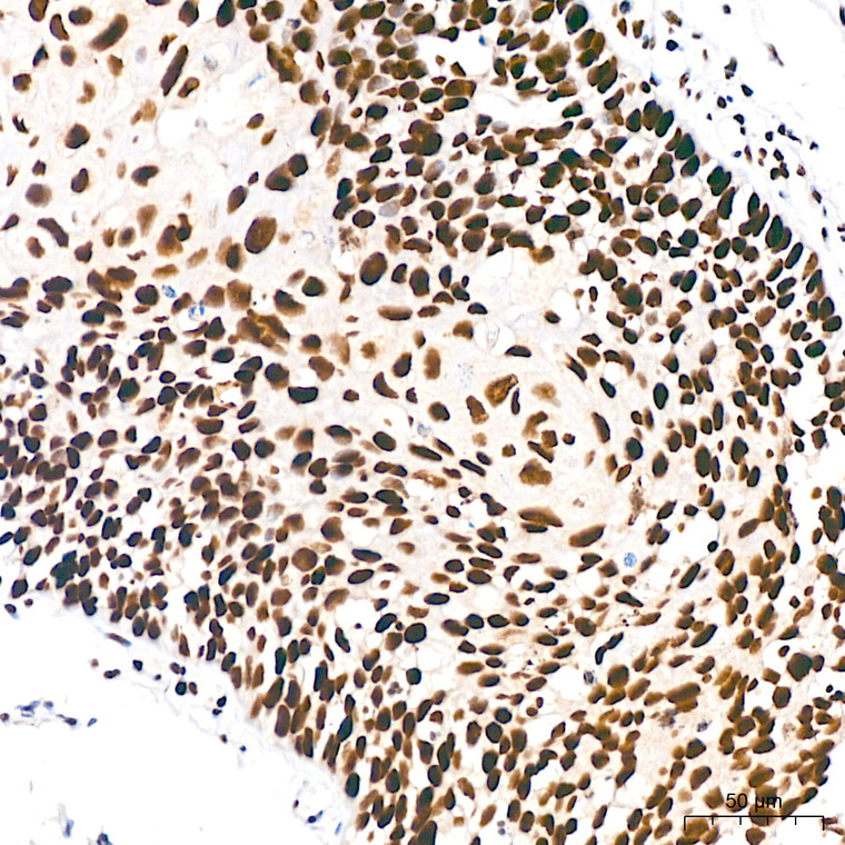
Immunohistochemistry analysis of DDX5 in paraffin-embedded human cervix cancer tissue using DDX5 rabbit monoclonal antibody (STJ11101314) at a dilution of 1:200 (40x lens). High pressure antigen retrieval was performed with 0. 01 M citrate buffer (pH 6. 0) prior to immunohistochemistry staining.
-
Immunohistochemistry analysis of DDX5 in paraffin-embedded human thyroid cancer tissue using DDX5 rabbit monoclonal antibody (STJ11101314) at a dilution of 1:200 (40x lens). High pressure antigen retrieval was performed with 0. 01 M citrate buffer (pH 6. 0) prior to immunohistochemistry staining.
-
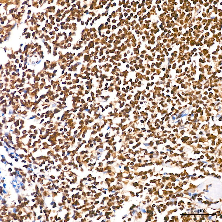
Immunohistochemistry analysis of DDX5 in paraffin-embedded human tonsil tissue using DDX5 rabbit monoclonal antibody (STJ11101314) at a dilution of 1:200 (40x lens). High pressure antigen retrieval was performed with 0. 01 M citrate buffer (pH 6. 0) prior to immunohistochemistry staining.
-
Immunohistochemistry analysis of DDX5 in paraffin-embedded mouse brain tissue using DDX5 rabbit monoclonal antibody (STJ11101314) at a dilution of 1:200 (40x lens). High pressure antigen retrieval was performed with 0. 01 M citrate buffer (pH 6. 0) prior to immunohistochemistry staining.
-
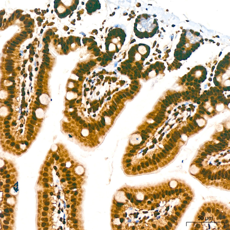
Immunohistochemistry analysis of DDX5 in paraffin-embedded mouse colon tissue using DDX5 rabbit monoclonal antibody (STJ11101314) at a dilution of 1:200 (40x lens). High pressure antigen retrieval was performed with 0. 01 M citrate buffer (pH 6. 0) prior to immunohistochemistry staining.
-
Immunohistochemistry analysis of DDX5 in paraffin-embedded mouse kidney tissue using DDX5 rabbit monoclonal antibody (STJ11101314) at a dilution of 1:200 (40x lens). High pressure antigen retrieval was performed with 0. 01 M citrate buffer (pH 6. 0) prior to immunohistochemistry staining.
-
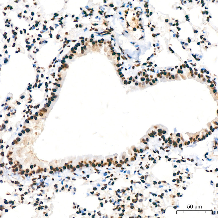
Immunohistochemistry analysis of DDX5 in paraffin-embedded mouse lung tissue using DDX5 rabbit monoclonal antibody (STJ11101314) at a dilution of 1:200 (40x lens). High pressure antigen retrieval was performed with 0. 01 M citrate buffer (pH 6. 0) prior to immunohistochemistry staining.
-
Immunohistochemistry analysis of DDX5 in paraffin-embedded mouse spleen tissue using DDX5 rabbit monoclonal antibody (STJ11101314) at a dilution of 1:200 (40x lens). High pressure antigen retrieval was performed with 0. 01 M citrate buffer (pH 6. 0) prior to immunohistochemistry staining.
-
Immunohistochemistry analysis of DDX5 in paraffin-embedded mouse testis tissue using DDX5 rabbit monoclonal antibody (STJ11101314) at a dilution of 1:200 (40x lens). High pressure antigen retrieval was performed with 0. 01 M citrate buffer (pH 6. 0) prior to immunohistochemistry staining.
-
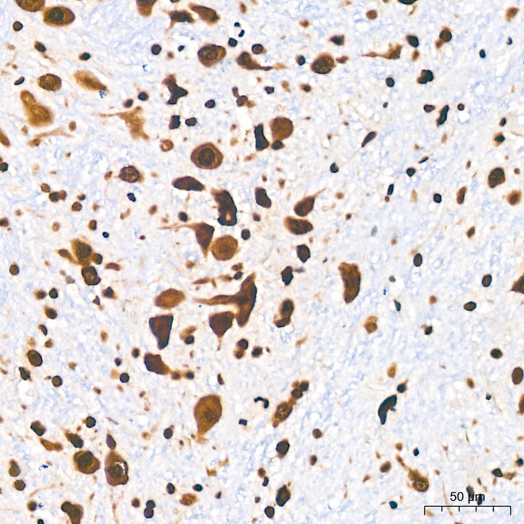
Immunohistochemistry analysis of DDX5 in paraffin-embedded rat brain tissue using DDX5 rabbit monoclonal antibody (STJ11101314) at a dilution of 1:200 (40x lens). High pressure antigen retrieval was performed with 0. 01 M citrate buffer (pH 6. 0) prior to immunohistochemistry staining.
-
Immunohistochemistry analysis of DDX5 in paraffin-embedded rat colon tissue using DDX5 rabbit monoclonal antibody (STJ11101314) at a dilution of 1:200 (40x lens). High pressure antigen retrieval was performed with 0. 01 M citrate buffer (pH 6. 0) prior to immunohistochemistry staining.
-
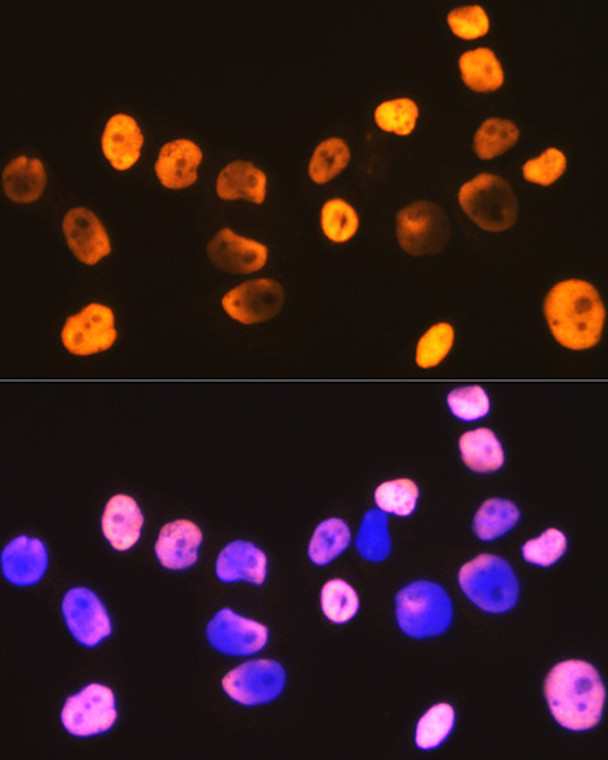
Immunofluorescence analysis of C6 cells using DDX5 rabbit monoclonal antibody (STJ11101314) at dilution of 1:100 (40x lens). Blue: DAPI for nuclear staining.
-
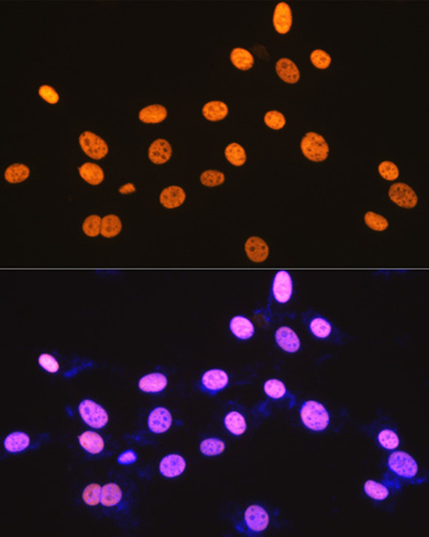
Immunofluorescence analysis of HeLa cells using DDX5 rabbit monoclonal antibody (STJ11101314) at dilution of 1:100 (40x lens). Blue: DAPI for nuclear staining.
-
Immunofluorescence analysis of NIH-3T3 cells using DDX5 rabbit monoclonal antibody (STJ11101314) at dilution of 1:100 (40x lens). Blue: DAPI for nuclear staining.
-
Immunoprecipitation analysis of 600 Mu g extracts of mouse testis using 3 Mu g DDX5 antibody (STJ11101314). Western blot was performed from the immunoprecipitate using DDX5 (STJ11101314) at a dilution of 1:1000.


