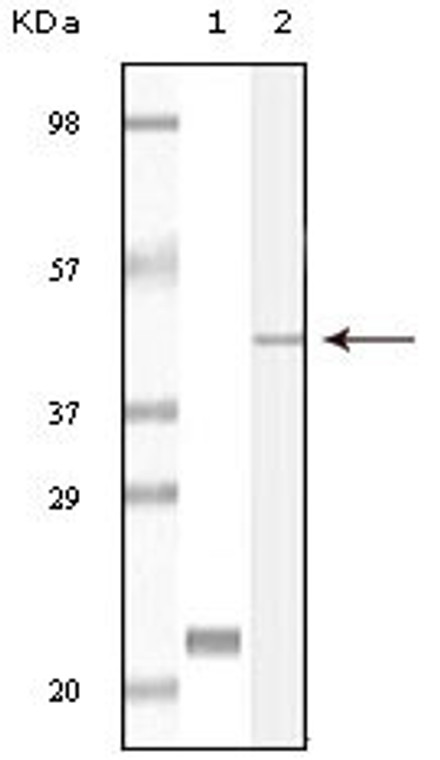
| Host: |
Mouse |
| Applications: |
WB/IHC/IF/ELISA |
| Reactivity: |
Human |
| Note: |
STRICTLY FOR FURTHER SCIENTIFIC RESEARCH USE ONLY (RUO). MUST NOT TO BE USED IN DIAGNOSTIC OR THERAPEUTIC APPLICATIONS. |
| Short Description: |
Mouse monoclonal antibody anti-Calcium and integrin-binding protein 1 is suitable for use in Western Blot, Immunohistochemistry, Immunofluorescence and ELISA research applications. |
| Clonality: |
Monoclonal |
| Clone ID: |
5A1F5E12/5A1H7E12 |
| Conjugation: |
Unconjugated |
| Formulation: |
Liquid in PBS containing 0.03% Sodium Azide, 0.5% BSA, 50% Glycerol. |
| Purification: |
Affinity purification |
| Dilution Range: |
WB 1:500-1:2000IHC 1:200-1:1000ELISA 1:10000IF 1:50-200 |
| Storage Instruction: |
Store at-20°C for up to 1 year from the date of receipt, and avoid repeat freeze-thaw cycles. |
| Gene Symbol: |
CIB1 |
| Gene ID: |
10519 |
| Uniprot ID: |
CIB1_HUMAN |
| Specificity: |
CIB1 Monoclonal Antibody detects endogenous levels of CIB1 protein. |
| Immunogen: |
Purified recombinant fragment of CIB1 expressed in E. Coli. |
| Post Translational Modifications | Phosphorylation of isoform 2 at Ser-118 by PRKD2 increases its ability to stimulate tumor angiogenesis. |
| Function | Calcium-binding protein that plays a role in the regulation of numerous cellular processes, such as cell differentiation, cell division, cell proliferation, cell migration, thrombosis, angiogenesis, cardiac hypertrophy and apoptosis. Involved in bone marrow megakaryocyte differentiation by negatively regulating thrombopoietin-mediated signaling pathway. Participates in the endomitotic cell cycle of megakaryocyte, a form of mitosis in which both karyokinesis and cytokinesis are interrupted. Plays a role in integrin signaling by negatively regulating alpha-IIb/beta3 activation in thrombin-stimulated megakaryocytes preventing platelet aggregation. Up-regulates PTK2/FAK1 activity, and is also needed for the recruitment of PTK2/FAK1 to focal adhesions.it thus appears to play an important role in focal adhesion formation. Positively regulates cell migration on fibronectin in a CDC42-dependent manner, the effect being negatively regulated by PAK1. Functions as a negative regulator of stress activated MAP kinase (MAPK) signaling pathways. Down-regulates inositol 1,4,5-trisphosphate receptor-dependent calcium signaling. Involved in sphingosine kinase SPHK1 translocation to the plasma membrane in a N-myristoylation-dependent manner preventing TNF-alpha-induced apoptosis. Regulates serine/threonine-protein kinase PLK3 activity for proper completion of cell division progression. Plays a role in microtubule (MT) dynamics during neuronal development.disrupts the MT depolymerization activity of STMN2 attenuating NGF-induced neurite outgrowth and the MT reorganization at the edge of lamellipodia. Promotes cardiomyocyte hypertrophy via activation of the calcineurin/NFAT signaling pathway. Stimulates calcineurin PPP3R1 activity by mediating its anchoring to the sarcolemma. In ischemia-induced (pathological or adaptive) angiogenesis, stimulates endothelial cell proliferation, migration and microvessel formation by activating the PAK1 and ERK1/ERK2 signaling pathway. Promotes also cancer cell survival and proliferation. May regulate cell cycle and differentiation of spermatogenic germ cells, and/or differentiation of supporting Sertoli cells. Isoform 2: Plays a regulatory role in angiogenesis and tumor growth by mediating PKD/PRKD2-induced vascular endothelial growth factor A (VEGFA) secretion. (Microbial infection) Involved in keratinocyte-intrinsic immunity to human beta-papillomaviruses (HPVs). |
| Protein Name | Calcium And Integrin-Binding Protein 1CibCalcium- And Integrin-Binding ProteinCibpCalmyrinDna-Pkcs-Interacting ProteinKinase-Interacting ProteinKipSnk-Interacting Protein 2-28Sip2-28 |
| Cellular Localisation | MembraneLipid-AnchorCell MembraneSarcolemmaApical Cell MembraneCell ProjectionRuffle MembraneFilopodium TipGrowth ConeLamellipodiumCytoplasmCytoskeletonMicrotubule Organizing CenterCentrosomePerinuclear RegionNucleusNeuron ProjectionPerikaryonColocalized With Ppp3r1 At The Cell Membrane Of Cardiomyocytes In The Hypertrophic HeartColocalized With Nbr1 To The Perinuclear RegionColocalizes With Tas1r2 In Apical Regions Of Taste Receptor CellsColocalized With Rac3 In The Perinuclear Area And At The Cell PeripheryColocalized With Pak1 Within Membrane Ruffles During Cell Spreading Upon Readhesion To FibronectinRedistributed To The Cytoskeleton Upon Platelet AggregationTranslocates From The Cytosol To The Plasma Membrane In A Calcium-Dependent MannerColocalized With Plk3 At Centrosomes In Ductal Breast Carcinoma CellsIsoform 2: CytoplasmGolgi ApparatusTrans-Golgi Network |
| Alternative Antibody Names | Anti-Calcium And Integrin-Binding Protein 1 antibodyAnti-Cib antibodyAnti-Calcium- And Integrin-Binding Protein antibodyAnti-Cibp antibodyAnti-Calmyrin antibodyAnti-Dna-Pkcs-Interacting Protein antibodyAnti-Kinase-Interacting Protein antibodyAnti-Kip antibodyAnti-Snk-Interacting Protein 2-28 antibodyAnti-Sip2-28 antibodyAnti-CIB1 antibodyAnti-CIB antibodyAnti-KIP antibodyAnti-PRKDCIP antibody |
Information sourced from Uniprot.org
12 months for antibodies. 6 months for ELISA Kits. Please see website T&Cs for further guidance


