| Host: |
Rabbit |
| Applications: |
WB |
| Reactivity: |
Human |
| Note: |
STRICTLY FOR FURTHER SCIENTIFIC RESEARCH USE ONLY (RUO). MUST NOT TO BE USED IN DIAGNOSTIC OR THERAPEUTIC APPLICATIONS. |
| Short Description: |
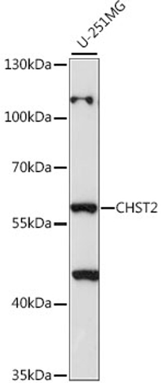
Rabbit polyclonal antibody anti-CHST2 (231-530) is suitable for use in Western Blot research applications. |
| Clonality: |
Polyclonal |
| Conjugation: |
Unconjugated |
| Isotype: |
IgG |
| Formulation: |
PBS with 0.01% Thimerosal, 50% Glycerol, pH7.3. |
| Purification: |
Affinity purification |
| Dilution Range: |
WB 1:500-1:2000 |
| Storage Instruction: |
Store at-20°C for up to 1 year from the date of receipt, and avoid repeat freeze-thaw cycles. |
| Gene Symbol: |
CHST2 |
| Gene ID: |
9435 |
| Uniprot ID: |
CHST2_HUMAN |
| Immunogen Region: |
231-530 |
| Immunogen: |
Recombinant fusion protein containing a sequence corresponding to amino acids 231-530 of human CHST2 (NP_004258.2). |
| Immunogen Sequence: |
FQLYSPAGSGGRNLTTLGIF GAATNKVVCSSPLCPAYRKE VVGLVDDRVCKKCPPQRLAR FEEECRKYRTLVIKGVRVFD VAVLAPLLRDPALDLKVIHL VRDPRAVASSRIRSRHGLIR ESLQVVRSRDPRAHRMPFLE AAGHKLGAKKEGVGGPADYH ALGAMEVICNSMAKTLQTAL QPPDWLQGHYLVVRYEDLVG DPVKTLRRVYDFVGLLVSPE MEQFALNMTSGSGSSSKPF |
| Tissue Specificity | Widely expressed. Highly expressed in bone marrow, peripheral blood leukocytes, spleen, brain, spinal cord, ovary and placenta. Expressed by high endothelial cells (HEVs) and leukocytes. |
| Post Translational Modifications | Glycosylation at Asn-475 is required for catalytic activity. |
| Function | Sulfotransferase that utilizes 3'-phospho-5'-adenylyl sulfate (PAPS) as sulfonate donor to catalyze the transfer of sulfate to position 6 of non-reducing N-acetylglucosamine (GlcNAc) residues within keratan-like structures on N-linked glycans and within mucin-associated glycans that can ultimately serve as SELL ligands. SELL ligands are present in high endothelial cells (HEVs) and play a central role in lymphocyte homing at sites of inflammation. Participates in biosynthesis of the SELL ligand sialyl 6-sulfo Lewis X and in lymphocyte homing to Peyer patches. Has no activity toward O-linked sugars. Its substrate specificity may be influenced by its subcellular location. Sulfates GlcNAc residues at terminal, non-reducing ends of oligosaccharide chains. |
| Protein Name | Carbohydrate Sulfotransferase 2Galactose/N-Acetylglucosamine/N-Acetylglucosamine 6-O-Sulfotransferase 2Gst-2N-Acetylglucosamine 6-O-Sulfotransferase 1Glcnac6st-1Gn6st-1 |
| Database Links | Reactome: R-HSA-2022854 |
| Cellular Localisation | Golgi ApparatusTrans-Golgi Network MembraneSingle-Pass Type Ii Membrane Protein |
| Alternative Antibody Names | Anti-Carbohydrate Sulfotransferase 2 antibodyAnti-Galactose/N-Acetylglucosamine/N-Acetylglucosamine 6-O-Sulfotransferase 2 antibodyAnti-Gst-2 antibodyAnti-N-Acetylglucosamine 6-O-Sulfotransferase 1 antibodyAnti-Glcnac6st-1 antibodyAnti-Gn6st-1 antibodyAnti-CHST2 antibodyAnti-GN6ST antibody |
Information sourced from Uniprot.org
12 months for antibodies. 6 months for ELISA Kits. Please see website T&Cs for further guidance


