| Host: |
Rabbit |
| Applications: |
WB/IF |
| Reactivity: |
Human/Mouse/Rat |
| Note: |
STRICTLY FOR FURTHER SCIENTIFIC RESEARCH USE ONLY (RUO). MUST NOT TO BE USED IN DIAGNOSTIC OR THERAPEUTIC APPLICATIONS. |
| Short Description: |
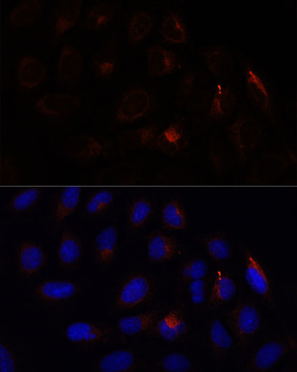
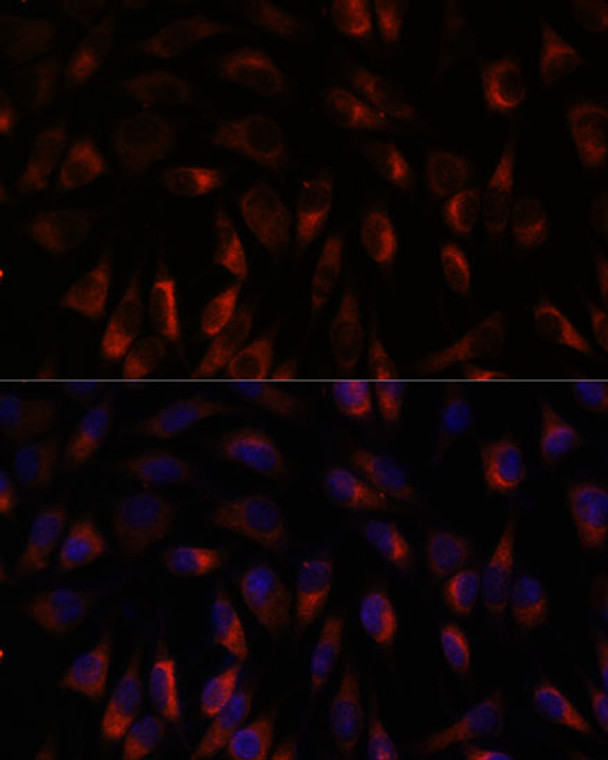
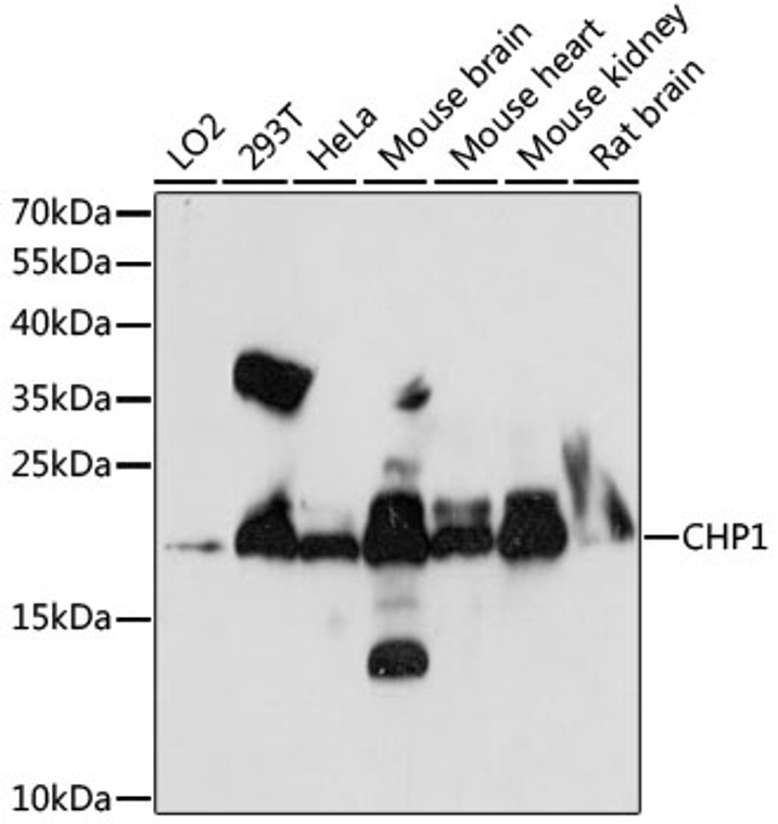
Rabbit polyclonal antibody anti-CHP1 (1-195) is suitable for use in Western Blot and Immunofluorescence research applications. |
| Clonality: |
Polyclonal |
| Conjugation: |
Unconjugated |
| Isotype: |
IgG |
| Formulation: |
PBS with 0.01% Thimerosal, 50% Glycerol, pH7.3. |
| Purification: |
Affinity purification |
| Dilution Range: |
WB 1:500-1:2000IF/ICC 1:50-1:200 |
| Storage Instruction: |
Store at-20°C for up to 1 year from the date of receipt, and avoid repeat freeze-thaw cycles. |
| Gene Symbol: |
CHP1 |
| Gene ID: |
11261 |
| Uniprot ID: |
CHP1_HUMAN |
| Immunogen Region: |
1-195 |
| Immunogen: |
Recombinant fusion protein containing a sequence corresponding to amino acids 1-195 of human CHP1 (NP_009167.1). |
| Immunogen Sequence: |
MGSRASTLLRDEELEEIKKE TGFSHSQITRLYSRFTSLDK GENGTLSREDFQRIPELAIN PLGDRIINAFFPEGEDQVNF RGFMRTLAHFRPIEDNEKSK DVNGPEPLNSRSNKLHFAFR LYDLDKDEKISRDELLQVLR MMVGVNISDEQLGSIADRTI QEADQDGDSAISFTEFVKVL EKVDVEQKMSIRFLH |
| Tissue Specificity | Ubiquitously expressed. Has been found in fetal eye, lung, liver, muscle, heart, kidney, thymus and spleen. |
| Post Translational Modifications | Phosphorylated.decreased phosphorylation is associated with an increase in SLC9A1/NHE1 Na(+)/H(+) exchange activity. Phosphorylation occurs in serum-dependent manner. The phosphorylation state may regulate the binding to SLC9A1/NHE1. Both N-myristoylation and calcium-mediated conformational changes are essential for its function in exocytic traffic. N-myristoylation is required for its association with microtubules and interaction with GAPDH, but not for the constitutive association to membranes. |
| Function | Calcium-binding protein involved in different processes such as regulation of vesicular trafficking, plasma membrane Na(+)/H(+) exchanger and gene transcription. Involved in the constitutive exocytic membrane traffic. Mediates the association between microtubules and membrane-bound organelles of the endoplasmic reticulum and Golgi apparatus and is also required for the targeting and fusion of transcytotic vesicles (TCV) with the plasma membrane. Functions as an integral cofactor in cell pH regulation by controlling plasma membrane-type Na(+)/H(+) exchange activity. Affects the pH sensitivity of SLC9A1/NHE1 by increasing its sensitivity at acidic pH. Required for the stabilization and localization of SLC9A1/NHE1 at the plasma membrane. Inhibits serum- and GTPase-stimulated Na(+)/H(+) exchange. Plays a role as an inhibitor of ribosomal RNA transcription by repressing the nucleolar UBF1 transcriptional activity. May sequester UBF1 in the nucleoplasm and limit its translocation to the nucleolus. Associates to the ribosomal gene promoter. Acts as a negative regulator of the calcineurin/NFAT signaling pathway. Inhibits NFAT nuclear translocation and transcriptional activity by suppressing the calcium-dependent calcineurin phosphatase activity. Also negatively regulates the kinase activity of the apoptosis-induced kinase STK17B. Inhibits both STK17B auto- and substrate-phosphorylations in a calcium-dependent manner. |
| Protein Name | Calcineurin B Homologous Protein 1Calcineurin B-Like ProteinCalcium-Binding Protein ChpCalcium-Binding Protein P22Ef-Hand Calcium-Binding Domain-Containing Protein P22 |
| Database Links | Reactome: R-HSA-2160916 |
| Cellular Localisation | NucleusCytoplasmCytoskeletonEndomembrane SystemEndoplasmic Reticulum-Golgi Intermediate CompartmentEndoplasmic ReticulumCell MembraneMembraneLipid-AnchorLocalizes In Cytoplasmic Compartments In Dividing CellsLocalizes In The Nucleus In Quiescent CellsExported From The Nucleus To The Cytoplasm Through A Nuclear Export Signal (Nes) And Crm1-Dependent PathwayMay Shuttle Between Nucleus And CytoplasmLocalizes With The Microtubule-Organizing Center (Mtoc) And Extends Toward The Periphery Along MicrotubulesAssociates With Membranes Of The Early Secretory Pathway In A Gapdh-IndependentN-Myristoylation- And Calcium-Dependent MannerColocalizes With The Mitotic Spindle MicrotubulesColocalizes With Gapdh Along MicrotubulesColocalizes With Slc9a1 At The Reticulum Endoplasmic And Plasma MembraneColocalizes With Stk17b At The Plasma Membrane |
| Alternative Antibody Names | Anti-Calcineurin B Homologous Protein 1 antibodyAnti-Calcineurin B-Like Protein antibodyAnti-Calcium-Binding Protein Chp antibodyAnti-Calcium-Binding Protein P22 antibodyAnti-Ef-Hand Calcium-Binding Domain-Containing Protein P22 antibodyAnti-CHP1 antibodyAnti-CHP antibody |
Information sourced from Uniprot.org
12 months for antibodies. 6 months for ELISA Kits. Please see website T&Cs for further guidance


