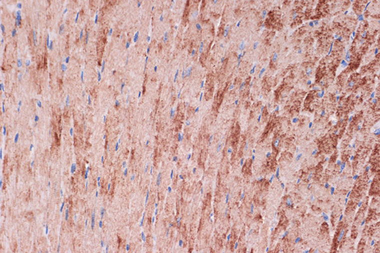
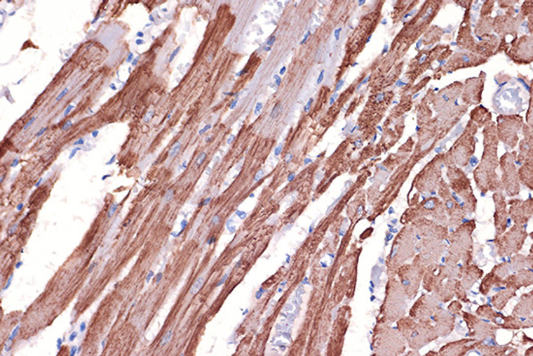
• Western blot analysis of extracts of various cell lines, using Tropomyosin 1 rabbit monoclonal antibody (STJ11102362) at 1:1000 dilution. Secondary antibody: HRP Goat Anti-rabbit IgG (H+L) at 1:10000 dilution. Lysates/proteins: 25ug per lane. Blocking buffer: 3% non-fat dry milk in TBST. Detection: ECL Basic Kit. Exposure time: 3s.
  • Immunoprecipitation analysis of 600 Mu g extracts of Rat heart using 3 Mu g Tropomyosin 1 Rabbit monoclonal antibody (STJ11102362). Western blot was performed from the immunoprecipitate using Tropomyosin 1 Rabbit monoclonal antibody (STJ11102362) at a dilution of 1:1000.
  • Immunohistochemistry of paraffin-embedded rat heart using Tropomyosin 1 rabbit monoclonal antibody (STJ11102362) at dilution of 1:100 (40x lens). Perform microwave antigen retrieval with 10 mM Tris/EDTA buffer pH 9. 0 before commencing with immunohistochemistry staining protocol.
  • Immunofluorescence analysis of paraffin-embedded mouse heart using Tropomyosin 1 Rabbit monoclonal antibody (STJ11102362) at dilution of 1:100 (40x lens). Secondary antibody: Cy3 Goat Anti-Rabbit IgG (H+L) at 1:500 dilution. Blue: DAPI for nuclear staining.
  • Immunohistochemistry of paraffin-embedded mouse heart using Tropomyosin 1 rabbit monoclonal antibody (STJ11102362) at dilution of 1:100 (40x lens). Perform microwave antigen retrieval with 10 mM Tris/EDTA buffer pH 9. 0 before commencing with immunohistochemistry staining protocol.
  • Immunofluorescence analysis of paraffin-embedded rat heart using Tropomyosin 1 Rabbit monoclonal antibody (STJ11102362) at dilution of 1:100 (40x lens). Secondary antibody: Cy3 Goat Anti-Rabbit IgG (H+L) at 1:500 dilution. Blue: DAPI for nuclear staining.
  • Immunofluorescence analysis of rat heart using Tropomyosin 1 rabbit monoclonal antibody (STJ11102362) at dilution of 1:100 (40x lens). Blue: DAPI for nuclear staining.
  • Immunohistochemistry analysis of Tropomyosin 1 in paraffin-embedded mouse heart using Tropomyosin 1 Rabbit monoclonal antibody (STJ11102362) at dilution of 1:100 (40x lens). Perform microwave antigen retrieval with 10 mM Tris/EDTA buffer pH 9. 0 before commencing with immunohistochemistry staining protocol.
  • Immunofluorescence analysis of mouse heart using Tropomyosin 1 rabbit monoclonal antibody (STJ11102362) at dilution of 1:100 (40x lens). Blue: DAPI for nuclear staining.
  • Immunohistochemistry analysis of Tropomyosin 1 in paraffin-embedded rat heart using Tropomyosin 1 Rabbit monoclonal antibody (STJ11102362) at dilution of 1:100 (40x lens). Perform microwave antigen retrieval with 10 mM Tris/EDTA buffer pH 9. 0 before commencing with immunohistochemistry staining protocol.
  • Immunoprecipitation analysis of 600ug extracts of Rat heart cells using 3ug Tropomyosin 1 antibody (STJ11102362). Western blot was performed from the immunoprecipitate using Tropomyosin 1 antibody (STJ11102362) at a dilition of 1:1000.
  • Western blot analysis of various lysates using Tropomyosin 1 Rabbit monoclonal antibody (STJ11102362) at 1:1000 dilution. Secondary antibody: HRP Goat Anti-Rabbit IgG (H+L) (STJS000856) at 1:10000 dilution. Lysates/proteins: 25 Mu g per lane. Blocking buffer: 3% nonfat dry milk in TBST. Detection: ECL Basic Kit. Exposure time: 3s.

Anti-TPM1 antibody (185-284) [S2MR] (STJ11102362)

SKU:
STJ11102362

Shipping:
Free Shipping
Current Stock:
Host: Rabbit
Applications: WB/IHC/IF/IP
Reactivity: Mouse/Rat
Note: STRICTLY FOR FURTHER SCIENTIFIC RESEARCH USE ONLY (RUO). MUST NOT TO BE USED IN DIAGNOSTIC OR THERAPEUTIC APPLICATIONS.
Short Description: Rabbit monoclonal antibody anti-Tropomyosin 1 (185-284) is suitable for use in Western Blot, Immunohistochemistry, Immunofluorescence and Immunoprecipitation research applications.
Clonality: Monoclonal
Clone ID: S2MR
Conjugation: Unconjugated
Isotype: IgG
Formulation: PBS with 0.02% Sodium Azide, 0.05% BSA, 50% Glycerol, pH7.3.
Purification: Affinity purification
Dilution Range: WB 1:500-1:2000
IHC-P 1:50-1:200
IF/ICC 1:50-1:200
IP 1:500-1:1000
Storage Instruction: Store at-20°C for up to 1 year from the date of receipt, and avoid repeat freeze-thaw cycles.
Gene Symbol: TPM1
Gene ID: 7168
Uniprot ID: TPM1_HUMAN
Immunogen Region: 185-284
Immunogen: A synthetic peptide corresponding to a sequence within amino acids 185-284 of human Tropomyosin 1 (P09493).
Immunogen Sequence: LSEGKCAELEEELKTVTNNL KSLEAQAEKYSQKEDRYEEE IKVLSDKLKEAETRAEFAER SVTKLEKSIDDLEDELYAQK LKYKAISEELDHALNDMTSI
Tissue Specificity Detected in primary breast cancer tissues but undetectable in normal breast tissues in Sudanese patients. Isoform 1 is expressed in adult and fetal skeletal muscle and cardiac tissues, with higher expression levels in the cardiac tissues. Isoform 10 is expressed in adult and fetal cardiac tissues, but not in skeletal muscle.
Post Translational Modifications Phosphorylated at Ser-283 by DAPK1 in response to oxidative stress and this phosphorylation enhances stress fiber formation in endothelial cells.
Function Binds to actin filaments in muscle and non-muscle cells. Plays a central role, in association with the troponin complex, in the calcium dependent regulation of vertebrate striated muscle contraction. Smooth muscle contraction is regulated by interaction with caldesmon. In non-muscle cells is implicated in stabilizing cytoskeleton actin filaments.
Protein Name Tropomyosin Alpha-1 Chain
Alpha-Tropomyosin
Tropomyosin-1
Database Links Reactome: R-HSA-390522
Reactome: R-HSA-445355
Cellular Localisation Cytoplasm
Cytoskeleton
Associates With F-Actin Stress Fibers
Alternative Antibody Names Anti-Tropomyosin Alpha-1 Chain antibody
Anti-Alpha-Tropomyosin antibody
Anti-Tropomyosin-1 antibody
Anti-TPM1 antibody
Anti-C15orf13 antibody
Anti-TMSA antibody

Information sourced from Uniprot.org

12 months for antibodies. 6 months for ELISA Kits. Please see website T&Cs for further guidance