


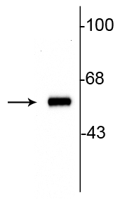
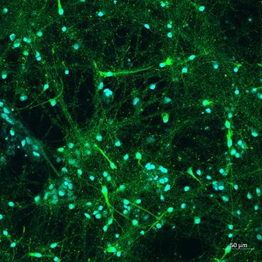
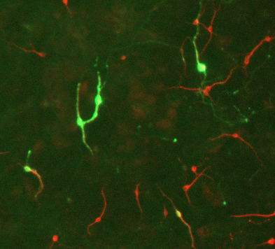
Anti-TH antibody (STJA0003816)
SPECIFICATIONS
ClonalityPolyclonal
HostRabbit
ConjugationUnconjugated
IsotypeIgG
ImmunogenSDS-denatured, native rat tyrosine hydroxylase purification from pheochromocytoma.
General Information
| Short Description | Rabbit polyclonal anti-Tyrosine Hydroxylase for use in WB, IHC and ICC in All Mammalian and Chicken samples. Datasheet included with dilution recommendations, and related reagents. |
| Applications | WB/IHC/ICC |
| Host | Rabbit |
| Reactivity | All Mammalian/Chicken |
| Note | STRICTLY FOR FURTHER SCIENTIFIC RESEARCH USE ONLY (RUO). MUST NOT TO BE USED IN DIAGNOSTIC OR THERAPEUTIC APPLICATIONS. |
Product Properties
| Clonality | Polyclonal |
| Isotype | IgG |
| Conjugation | Unconjugated |
| Purification | This antibody was antigen affinity purified from pooled serum. |
| Dilution Range | WB 1:1000IHC 1:1000ICC 1:500-1:1000 |
| Formulation | 100 µl in 10 mM HEPES (pH 7.5) , 150 mM NaCl, 100 µg per ml BSA and 50% Glycerol. |
| Storage Instruction | Store at-20°C for up to 1 year from the date of receipt, and avoid repeat freeze-thaw cycles. |
Target Information
| Gene Symbol | Th |
| Gene ID | 25085 |
| Uniprot ID | TY3H_RAT |
| Immunogen | SDS-denatured, native rat tyrosine hydroxylase purification from pheochromocytoma. |
Additional Info
| Post Translational Modifications | Phosphorylated on Ser-19, Ser-31 and Ser-40 by several protein kinases with different site specificities. Phosphorylation at Ser-31 and Ser-40 leads to an increase of TH activity. Phosphorylation at Ser-40 activates the enzyme and also counteracts the feedback inhibition of TH by catecholamines. Phosphorylation of Ser-19 and Ser-31 triggers the proteasomal degradation of TH through the ubiquitin-proteasome pathway. Phosphorylation at Ser-31 facilitates transport of TH from the soma to the nerve terminals via the microtubule network. Phosphorylation at Ser-19 induces the high-affinity binding to the 14-3-3 protein YWHAG.this interaction may influence the phosphorylation and dephosphorylation of other sites. Ser-19 increases the phosphorylation at Ser-40 in a hierarchical manner, leading to increased activity. |
| Function | Catalyzes the conversion of L-tyrosine to L-dihydroxyphenylalanine (L-Dopa), the rate-limiting step in the biosynthesis of catecholamines, dopamine, noradrenaline, and adrenaline. Uses tetrahydrobiopterin and molecular oxygen to convert tyrosine to L-Dopa. In addition to tyrosine, is able to catalyze the hydroxylation of phenylalanine and tryptophan but with lower specificity. Positively regulates the regression of retinal hyaloid vessels during postnatal development. |
| Protein Name | Tyrosine 3-MonooxygenaseTyrosine 3-HydroxylaseTh |
| Database Links | Reactome: R-RNO-209905 |
| Cellular Localisation | CytoplasmPerinuclear RegionNucleusCell ProjectionAxonCytoplasmic VesicleSecretory VesicleSynaptic VesicleWhen Phosphorylated At Ser-19 Shows A Nuclear Distribution And When Phosphorylated At Ser-31 As Well As At Ser-40 Shows A Cytosolic DistributionExpressed In Dopaminergic Axons And Axon Terminals |
| Alternative Antibody Names | Anti-Tyrosine 3-Monooxygenase antibodyAnti-Tyrosine 3-Hydroxylase antibodyAnti-Th antibodyAnti-Th antibody |
Information sourced from Uniprot.org