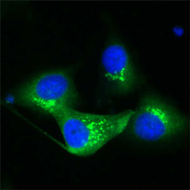
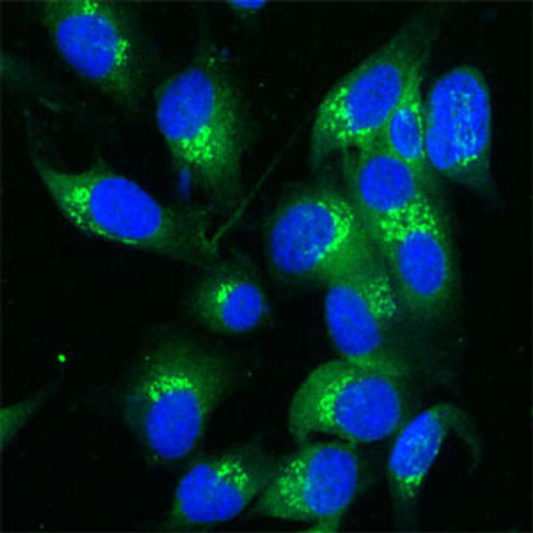
• Human Melanoma cell line C 32 was cultured overnight on round cover slides placed in a 24 well tissue culture plate. Culture media removed and washed twice with PBS before fixing with 2% formalin for 10 minutes. Cells were then washed three times with PBS and incubated with Tris 0. 01M containing Triton X 0. 005% for 15 minutes. Cells were washed and incubated with 100 µl of Rabbit antibody to TGN (STJ13100506) diluted 1:100 in the blocking buffer for 30 minutes. Welles were then washed 7 times with PBS and incubated with 100 µl of anti Rb-FITC conjugate diluted 1:100 in the blocking buffer for further 30 minutes. Cells were washed as before and nuclear counter stained with Hoechst and mounted on to slides.
  • Human Melanoma cell line C 32 was cultured overnight on round cover slides placed in a 24 well tissue culture plate. Culture media removed and washed twice with PBS before fixing with 2% formalin for 10 minutes. Cells were then washed three times with PBS and incubated with Tris 0. 01M containing Triton X 0. 005% for 15 minutes. Cells were washed and incubated with 100 µl of Rabbit antibody to TGN (STJ13100506) diluted 1:100 in the blocking buffer for 30 minutes. Welles were then washed 7 times with PBS and incubated with 100 µl of anti Rb-FITC conjugate diluted 1:100 in the blocking buffer for further 30 minutes. Cells were washed as before and nuclear counter stained with Hoechst and mounted on to slides.
  • Human Melanoma cell line C 32 was cultured overnight on round cover slides placed in a 24 well tissue culture plate. Culture media removed and washed twice with PBS before fixing with 2% formalin for 10 minutes. Cells were then washed three times with PBS and incubated with Tris 0. 01M containing Triton X 0. 005% for 15 minutes. Cells were washed and incubated with 100 µl of Rabbit antibody to TGN (STJ13100506) diluted 1:100 in the blocking buffer for 30 minutes. Welles were then washed 7 times with PBS and incubated with 100 µl of anti Rb-FITC conjugate diluted 1:100 in the blocking buffer for further 30 minutes. Cells were washed as before and nuclear counter stained with Hoechst and mounted on to slides.
  • Human Melanoma cell line C 32 was cultured overnight on round cover slides placed in a 24 well tissue culture plate. Culture media removed and washed twice with PBS before fixing with 2% formalin for 10 minutes. Cells were then washed three times with PBS and incubated with Tris 0. 01M containing Triton X 0. 005% for 15 minutes. Cells were washed and incubated with 100 µl of Rabbit antibody to TGN (STJ13100506) diluted 1:100 in the blocking buffer for 30 minutes. Welles were then washed 7 times with PBS and incubated with 100 µl of anti Rb-FITC conjugate diluted 1:100 in the blocking buffer for further 30 minutes. Cells were washed as before and nuclear counter stained with Hoechst and mounted on to slides.

Anti-TGN38 antibody (STJ13100506)

SKU:
STJ13100506-500

Shipping:
Free Shipping
Current Stock:
Host: NZ White Rabbit
Applications: IHC/WB
Reactivity: Rat/Mouse/Human/Marmoset
Note: STRICTLY FOR FURTHER SCIENTIFIC RESEARCH USE ONLY (RUO). MUST NOT TO BE USED IN DIAGNOSTIC OR THERAPEUTIC APPLICATIONS.
Short Description: Nz White Rabbit polyclonal antibody anti-TGN38 is suitable for use in Immunohistochemistry and Western Blot research applications.
Clonality: Polyclonal
Conjugation: Unconjugated
Isotype: IgG
Formulation: Shipped as lyophilised. Reconstitute in 500 µl of sterile water. Centrifuge to remove any insoluble material.
Purification: Ammonium sulphate precipitation
Dilution Range: A concentration of 10-50 µg/ml is recommended. The optimal concentration should be determined by the end user. Not yet tested in other applications.
Storage Instruction: Maintain the lyophilised/reconstituted antibodies frozen at-20°C for long term storage and refrigerated at 2-8°C for a shorter term. When reconstituting, glycerol (1:1) may be added for an additional stability. Avoid freeze and thaw cycles.
Gene Symbol: Tgoln2
Gene ID: 22135
Uniprot ID: TGON2_MOUSE
Specificity: Specific for TGN38 (or TGN46, TGN48, TGN51 in human).
Immunogen: A synthetic peptide from mouse TGN38 conjugated to blue carrier protein was used as the antigen.
Function May be involved in regulating membrane traffic to and from trans-Golgi network.
Protein Name Trans-Golgi Network Integral Membrane Protein 2
Tgn38b
Cellular Localisation Cell Membrane
Single-Pass Type I Membrane Protein
Golgi Apparatus
Trans-Golgi Network Membrane
Primarily In Trans-Golgi Network
Cycles Between The Trans-Golgi Network And The Cell Surface Returning Via Endosomes
Alternative Antibody Names Anti-Trans-Golgi Network Integral Membrane Protein 2 antibody
Anti-Tgn38b antibody
Anti-Tgoln2 antibody
Anti-Ttgn2 antibody

Information sourced from Uniprot.org

12 months for antibodies. 6 months for ELISA Kits. Please see website T&Cs for further guidance