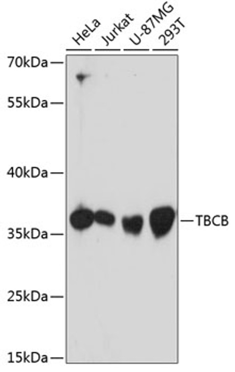
| Host: |
Rabbit |
| Applications: |
WB |
| Reactivity: |
Human |
| Note: |
STRICTLY FOR FURTHER SCIENTIFIC RESEARCH USE ONLY (RUO). MUST NOT TO BE USED IN DIAGNOSTIC OR THERAPEUTIC APPLICATIONS. |
| Short Description: |
Rabbit polyclonal antibody anti-TBCB (1-244) is suitable for use in Western Blot research applications. |
| Clonality: |
Polyclonal |
| Conjugation: |
Unconjugated |
| Isotype: |
IgG |
| Formulation: |
PBS with 0.01% Thimerosal, 50% Glycerol, pH7.3. |
| Purification: |
Affinity purification |
| Dilution Range: |
WB 1:500-1:2000 |
| Storage Instruction: |
Store at-20°C for up to 1 year from the date of receipt, and avoid repeat freeze-thaw cycles. |
| Gene Symbol: |
TBCB |
| Gene ID: |
1155 |
| Uniprot ID: |
TBCB_HUMAN |
| Immunogen Region: |
1-244 |
| Immunogen: |
Recombinant fusion protein containing a sequence corresponding to amino acids 1-244 of human TBCB (NP_001272.2). |
| Immunogen Sequence: |
MEVTGVSAPTVTVFISSSLN TFRSEKRYSRSLTIAEFKCK LELLVGSPASCMELELYGVD DKFYSKLDQEDALLGSYPVD DGCRIHVIDHSGARLGEYED VSRVEKYTISQEAYDQRQDT VRSFLKRSKLGRYNEEERAQ QEAEAAQRLAEEKAQASSIP VGSRCEVRAAGQSPRRGTVM YVGLTDFKPGYWIGVRYDEP LGKNDGSVNGKRYFECQAKY GAFVKPAVVTVGDFPEEDY |
| Tissue Specificity | Found in most tissues. |
| Post Translational Modifications | Phosphorylation by PAK1 is required for normal function. Ubiquitinated in the presence of GAN which targets it for degradation by the proteasome. (Microbial infection) Glycosylated residues by S.typhimurium protein Ssek1: arginine GlcNAcylation promotes microtubule stability. |
| Function | Binds to alpha-tubulin folding intermediates after their interaction with cytosolic chaperonin in the pathway leading from newly synthesized tubulin to properly folded heterodimer. Involved in regulation of tubulin heterodimer dissociation. May function as a negative regulator of axonal growth. |
| Protein Name | Tubulin-Folding Cofactor BCytoskeleton-Associated Protein 1Cytoskeleton-Associated Protein CkapiTubulin-Specific Chaperone B |
| Database Links | Reactome: R-HSA-389977 |
| Cellular Localisation | CytoplasmCytoskeletonColocalizes With MicrotubulesIn Differentiated NeuronsLocated In The CytoplasmIn Differentiating NeuronsAccumulates At The Growth Cone |
| Alternative Antibody Names | Anti-Tubulin-Folding Cofactor B antibodyAnti-Cytoskeleton-Associated Protein 1 antibodyAnti-Cytoskeleton-Associated Protein Ckapi antibodyAnti-Tubulin-Specific Chaperone B antibodyAnti-TBCB antibodyAnti-CG22 antibodyAnti-CKAP1 antibody |
Information sourced from Uniprot.org
12 months for antibodies. 6 months for ELISA Kits. Please see website T&Cs for further guidance


![Western blot analysis of lysates from wild type (WT) and LRRC59 knockout (KO) 293T cells, using [KO Validated] LRRC59 Rabbit polyclonal antibody (STJ11100841) at 1:1000 dilution. Secondary antibody: HRP Goat Anti-Rabbit IgG (H+L) (STJS000856) at 1:10000 dilution. Lysates/proteins: 25 Mu g per lane. Blocking buffer: 3% nonfat dry milk in TBST. Detection: ECL Basic Kit. Exposure time: 5s. Western blot analysis of lysates from wild type (WT) and LRRC59 knockout (KO) 293T cells, using [KO Validated] LRRC59 Rabbit polyclonal antibody (STJ11100841) at 1:1000 dilution. Secondary antibody: HRP Goat Anti-Rabbit IgG (H+L) (STJS000856) at 1:10000 dilution. Lysates/proteins: 25 Mu g per lane. Blocking buffer: 3% nonfat dry milk in TBST. Detection: ECL Basic Kit. Exposure time: 5s.](https://cdn11.bigcommerce.com/s-zso2xnchw9/images/stencil/300x300/products/89775/358247/STJ11100841_1__11256.1713122582.jpg?c=1)