


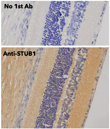
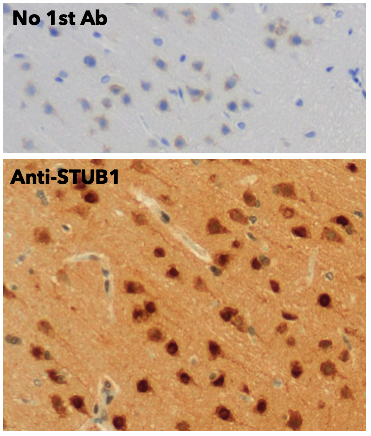
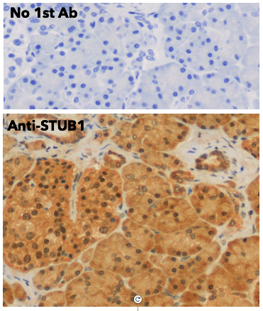
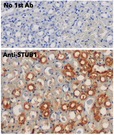
Anti-STUB1 antibody (STJ140143)
SPECIFICATIONS
ClonalityPolyclonal
HostGoat
ConjugationUnconjugated
IsotypeIgG
ImmunogenPurified recombinant human STUB1 protein produced in E. coli.
General Information
| Short Description | Goat polyclonal STIP1 homology and U-box containing protein 1, E3 ubiquitin protein ligase antibody for use in WB, IHC-P and IHC-F in human, mouse, rat, bovine, canine, chicken, avian, donkey, feline, goat, guinea pig, hamster, horse, porcine, rabbit |
| Applications | WB/IHC-P/IHC-F |
| Host | Goat |
| Reactivity | Human/Mouse/Rat/Bovine/Canine/Chicken/Avian/Donkey/Feline/Goat/Guinea Pig/Hamster/Horse/Porcine/Rabbit/Sheep/Simian |
| Note | STRICTLY FOR FURTHER SCIENTIFIC RESEARCH USE ONLY (RUO). MUST NOT TO BE USED IN DIAGNOSTIC OR THERAPEUTIC APPLICATIONS. |
Product Properties
| Clonality | Polyclonal |
| Isotype | IgG |
| Conjugation | Unconjugated |
| Concentration | 3 mg/mL |
| Purification | This antibody is epitope-affinity purified from goat antiserum. |
| Dilution Range | WB 1:500-1:5000IHC-F 1:250-1:1000IHC-P 1:250-1:1000 |
| Formulation | PBS, 20% Glycerol and 0.05% Sodium Azide. |
| Storage Instruction | For continuous use, store at 2-8 C for one-two days. For extended storage, store in-20 C freezer. Working dilution samples should be discarded if not used within 12 hours. |
Target Information
| Gene Symbol | STUB1 |
| Gene ID | 10273 |
| Uniprot ID | CHIP_HUMAN |
| Accession Number | ENSG00000103266 |
| Immunogen | Purified recombinant human STUB1 protein produced in E. coli. |
| Immunogen Sequence | MKGKEEKEGGARLGAGGGSP KGKEEKEGGARLGAGGGSPE GKEEKEGGARLGAGGGSPEK KEEKEGGARLGAGGGSPEKS EEKEGGARLGAGGGSPEKSP EKEGGARLGAGGGSPEKSPS KEGGARLGAGGGSPEKSPSA EGGARLGAGGGSPEKSPSAQ GGARLGAGGGSPEKSPSAQE GARLGAGGGSPEKSPSAQEL ARLGAGGGSPEKSPSAQELK RLGAGGGSPEKSPSAQELK |
| Specificity | Detects endogenous levels of STUB1 by Western blot. |
Additional Info
| Tissue Specificity | Expressed in differentiated myotubes (at protein level). Highly expressed in skeletal muscle, heart, pancreas, brain and placenta. Detected in kidney, liver and lung. |
| Post Translational Modifications | Monoubiquitinated at Lys-2 following cell stress by UBE2W, promoting the interaction with ATXN3. Auto-ubiquitinated.mediated by UBE2D1 and UBE2D2 and enhanced in the presence of MAP2K5. |
| Function | E3 ubiquitin-protein ligase which targets misfolded chaperone substrates towards proteasomal degradation. Plays a role in the maintenance of mitochondrial morphology and promotes mitophagic removal of dysfunctional mitochondria.thereby acts as a protector against apoptosis in response to cellular stress. Negatively regulates vascular smooth muscle contraction, via degradation of the transcriptional activator MYOCD and subsequent loss of transcription of genes involved in vascular smooth muscle contraction. Promotes survival and proliferation of cardiac smooth muscle cells via ubiquitination and degradation of FOXO1, resulting in subsequent repression of FOXO1-mediated transcription of pro-apoptotic genes. Ubiquitinates ICER-type isoforms of CREM and targets them for proteasomal degradation, thereby acts as a positive effector of MAPK/ERK-mediated inhibition of apoptosis in cardiomyocytes. Inhibits lipopolysaccharide-induced apoptosis and hypertrophy in cardiomyocytes, via ubiquitination and subsequent proteasomal degradation of NFATC3. Collaborates with ATXN3 in the degradation of misfolded chaperone substrates: ATXN3 restricting the length of ubiquitin chain attached to STUB1/CHIP substrates and preventing further chain extension. Ubiquitinates NOS1 in concert with Hsp70 and Hsp40. Modulates the activity of several chaperone complexes, including Hsp70, Hsc70 and Hsp90. Ubiquitinates CHRNA3 targeting it for endoplasmic reticulum-associated degradation in cortical neurons, as part of the STUB1-VCP-UBXN2A complex. Ubiquitinates and promotes ESR1 proteasomal degradation in response to age-related circulating estradiol (17-beta-estradiol/E2) decline, thereby promotes neuronal apoptosis in response to ischemic reperfusion injury. Mediates transfer of non-canonical short ubiquitin chains to HSPA8 that have no effect on HSPA8 degradation. Mediates polyubiquitination of DNA polymerase beta (POLB) at 'Lys-41', 'Lys-61' and 'Lys-81', thereby playing a role in base-excision repair: catalyzes polyubiquitination by amplifying the HUWE1/ARF-BP1-dependent monoubiquitination and leading to POLB-degradation by the proteasome. Mediates polyubiquitination of CYP3A4. Ubiquitinates EPHA2 and may regulate the receptor stability and activity through proteasomal degradation. Acts as a co-chaperone for HSPA1A and HSPA1B chaperone proteins and promotes ubiquitin-mediated protein degradation. Negatively regulates the suppressive function of regulatory T-cells (Treg) during inflammation by mediating the ubiquitination and degradation of FOXP3 in a HSPA1A/B-dependent manner. Catalyzes monoubiquitination of SIRT6, preventing its degradation by the proteasome. Likely mediates polyubiquitination and down-regulates plasma membrane expression of PD-L1/CD274, an immune inhibitory ligand critical for immune tolerance to self and antitumor immunity. Negatively regulates TGF-beta signaling by modulating the basal level of SMAD3 via ubiquitin-mediated degradation. Plays a role in the degradation of TP53. Mediates ubiquitination of RIPK3 leading to its subsequent proteasome-dependent degradation. May regulate myosin assembly in striated muscles together with UBE4B and VCP/p97 by targeting myosin chaperone UNC45B for proteasomal degradation. Ubiquitinates PPARG in macrophages playing a role in M2 macrophages polarization and angiogenesis. |
| Protein Name | E3 Ubiquitin-Protein Ligase ChipAntigen Ny-Co-7Cll-Associated Antigen Kw-8Carboxy Terminus Of Hsp70-Interacting ProteinRing-Type E3 Ubiquitin Transferase ChipStip1 Homology And U Box-Containing Protein 1 |
| Database Links | Reactome: R-HSA-2173788Reactome: R-HSA-5213460Reactome: R-HSA-5357905Reactome: R-HSA-5675482Reactome: R-HSA-8863795Reactome: R-HSA-8939902Reactome: R-HSA-8948751Reactome: R-HSA-983168 |
| Cellular Localisation | CytoplasmNucleusMitochondrionTranslocates To The Nucleus In Response To Inflammatory Signals In Regulatory T-Cells (Treg)Localizes To Mitochondria Following Oxygen And Glucose Deprivation-Induced Cellular Stress |
| Alternative Antibody Names | Anti-E3 Ubiquitin-Protein Ligase Chip antibodyAnti-Antigen Ny-Co-7 antibodyAnti-Cll-Associated Antigen Kw-8 antibodyAnti-Carboxy Terminus Of Hsp70-Interacting Protein antibodyAnti-Ring-Type E3 Ubiquitin Transferase Chip antibodyAnti-Stip1 Homology And U Box-Containing Protein 1 antibodyAnti-STUB1 antibodyAnti-CHIP antibodyAnti-PP1131 antibody |
Information sourced from Uniprot.org