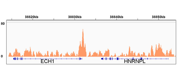
| Host: |
Rabbit |
| Applications: |
WB/IF/IP |
| Reactivity: |
Human/Mouse/Rat |
| Note: |
STRICTLY FOR FURTHER SCIENTIFIC RESEARCH USE ONLY (RUO). MUST NOT TO BE USED IN DIAGNOSTIC OR THERAPEUTIC APPLICATIONS. |
| Short Description: |
Rabbit monoclonal antibody anti-RXR Alpha (1-100) is suitable for use in Western Blot, Immunofluorescence and Immunoprecipitation research applications. |
| Clonality: |
Monoclonal |
| Clone ID: |
S2MR |
| Conjugation: |
Unconjugated |
| Isotype: |
IgG |
| Formulation: |
PBS with 0.02% Sodium Azide, 0.05% BSA, 50% Glycerol, pH7.3. |
| Purification: |
Affinity purification |
| Dilution Range: |
WB 1:500-1:2000IF/ICC 1:50-1:200IP 1:500-1:1000ChIP 1:500-1:1000 |
| Storage Instruction: |
Store at-20°C for up to 1 year from the date of receipt, and avoid repeat freeze-thaw cycles. |
| Gene Symbol: |
RXRA |
| Gene ID: |
6256 |
| Uniprot ID: |
RXRA_HUMAN |
| Immunogen Region: |
1-100 |
| Immunogen: |
A synthetic peptide corresponding to a sequence within amino acids 1-100 of human RXR Alpha (P19793). |
| Immunogen Sequence: |
MDTKHFLPLDFSTQVNSSLT SPTGRGSMAAPSLHPSLGPG IGSPGQLHSPISTLSSPING MGPPFSVISSPMGPHSMSVP TTPTLGFSTGSPQLSSPMNP |
| Tissue Specificity | Expressed in lung fibroblasts (at protein level). Expressed in monocytes. Highly expressed in liver, also found in kidney and brain. |
| Post Translational Modifications | Acetylated by EP300.acetylation enhances DNA binding and transcriptional activity. Phosphorylated on serine and threonine residues mainly in the N-terminal modulating domain. Constitutively phosphorylated on Ser-21 in the presence or absence of ligand. Under stress conditions, hyperphosphorylated by activated JNK on Ser-56, Ser-70, Thr-82 and Ser-260. Phosphorylated on Ser-27, in vitro, by PKA. This phosphorylation is required for repression of cAMP-mediated transcriptional activity of RARA. Sumoylation negatively regulates transcriptional activity. Desumoylated specifically by SENP6. |
| Function | Receptor for retinoic acid that acts as a transcription factor. Forms homo- or heterodimers with retinoic acid receptors (RARs) and binds to target response elements in response to their ligands, all-trans or 9-cis retinoic acid, to regulate gene expression in various biological processes. The RAR/RXR heterodimers bind to the retinoic acid response elements (RARE) composed of tandem 5'-AGGTCA-3' sites known as DR1-DR5 to regulate transcription. The high affinity ligand for retinoid X receptors (RXRs) is 9-cis retinoic acid. In the absence of ligand, the RXR-RAR heterodimers associate with a multiprotein complex containing transcription corepressors that induce histone deacetylation, chromatin condensation and transcriptional suppression. On ligand binding, the corepressors dissociate from the receptors and coactivators are recruited leading to transcriptional activation. Serves as a common heterodimeric partner for a number of nuclear receptors, such as RARA, RARB and PPARA. The RXRA/RARB heterodimer can act as a transcriptional repressor or transcriptional activator, depending on the RARE DNA element context. The RXRA/PPARA heterodimer is required for PPARA transcriptional activity on fatty acid oxidation genes such as ACOX1 and the P450 system genes. Together with RARA, positively regulates microRNA-10a expression, thereby inhibiting the GATA6/VCAM1 signaling response to pulsatile shear stress in vascular endothelial cells. Acts as an enhancer of RARA binding to RARE DNA element. May facilitate the nuclear import of heterodimerization partners such as VDR and NR4A1. Promotes myelin debris phagocytosis and remyelination by macrophages. Plays a role in the attenuation of the innate immune system in response to viral infections, possibly by negatively regulating the transcription of antiviral genes such as type I IFN genes. Involved in the regulation of calcium signaling by repressing ITPR2 gene expression, thereby controlling cellular senescence. |
| Protein Name | Retinoic Acid Receptor Rxr-AlphaNuclear Receptor Subfamily 2 Group B Member 1Retinoid X Receptor Alpha |
| Database Links | Reactome: R-HSA-1368082Reactome: R-HSA-1368108Reactome: R-HSA-159418Reactome: R-HSA-192105Reactome: R-HSA-193368Reactome: R-HSA-193807Reactome: R-HSA-1989781Reactome: R-HSA-200425Reactome: R-HSA-204174Reactome: R-HSA-211976Reactome: R-HSA-2151201Reactome: R-HSA-2426168Reactome: R-HSA-381340Reactome: R-HSA-383280Reactome: R-HSA-400206Reactome: R-HSA-400253Reactome: R-HSA-4090294Reactome: R-HSA-5362517Reactome: R-HSA-5617472Reactome: R-HSA-9029558Reactome: R-HSA-9029569Reactome: R-HSA-9031525Reactome: R-HSA-9031528Reactome: R-HSA-9616222Reactome: R-HSA-9623433Reactome: R-HSA-9632974Reactome: R-HSA-9707564Reactome: R-HSA-9707616 |
| Cellular Localisation | NucleusCytoplasmMitochondrionLocalization To The Nucleus Is Enhanced By Vitamin D3Nuclear Localization May Be Enhanced By The Interaction With Heterodimerization Partner VdrTranslocation To The Mitochondrion Upon Interaction With Nr4a1Increased Nuclear Localization Upon Pulsatile Shear Stress |
| Alternative Antibody Names | Anti-Retinoic Acid Receptor Rxr-Alpha antibodyAnti-Nuclear Receptor Subfamily 2 Group B Member 1 antibodyAnti-Retinoid X Receptor Alpha antibodyAnti-RXRA antibodyAnti-NR2B1 antibody |
Information sourced from Uniprot.org
12 months for antibodies. 6 months for ELISA Kits. Please see website T&Cs for further guidance


