| Host: |
Rabbit |
| Applications: |
WB |
| Reactivity: |
Human/Rat |
| Note: |
STRICTLY FOR FURTHER SCIENTIFIC RESEARCH USE ONLY (RUO). MUST NOT TO BE USED IN DIAGNOSTIC OR THERAPEUTIC APPLICATIONS. |
| Short Description: |
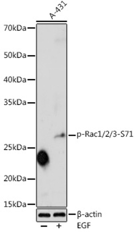
Rabbit polyclonal antibody anti-Phospho-RAC1-S71 is suitable for use in Western Blot research applications. |
| Clonality: |
Polyclonal |
| Conjugation: |
Unconjugated |
| Isotype: |
IgG |
| Formulation: |
PBS with 0.01% Thimerosal, 50% Glycerol, pH7.3. |
| Purification: |
Affinity purification |
| Dilution Range: |
WB 1:500-1:1000 |
| Storage Instruction: |
Store at-20°C for up to 1 year from the date of receipt, and avoid repeat freeze-thaw cycles. |
| Gene Symbol: |
RAC1 |
| Gene ID: |
5879 |
| Uniprot ID: |
RAC1_HUMAN |
| Immunogen: |
A synthetic phosphorylated peptide around S71 of human RAC1 (NP_008839.2). |
| Immunogen Sequence: |
YDRLRPLSYPQTDVF |
| Tissue Specificity | Isoform B is predominantly identified in skin and epithelial tissues from the intestinal tract. Its expression is elevated in colorectal tumors at various stages of neoplastic progression, as compared to their respective adjacent tissues. |
| Post Translational Modifications | GTP-bound active form is ubiquitinated by HACE1, leading to its degradation by the proteasome. Phosphorylated by AKT at Ser-71. Ubiquitinated at Lys-166 in a FBXL19-mediated manner.leading to proteasomal degradation. (Microbial infection) AMPylation at Tyr-32 and Thr-35 are mediated by bacterial enzymes in case of infection by H.somnus and V.parahaemolyticus, respectively. AMPylation occurs in the effector region and leads to inactivation of the GTPase activity by preventing the interaction with downstream effectors, thereby inhibiting actin assembly in infected cells. It is unclear whether some human enzyme mediates AMPylation.FICD has such ability in vitro but additional experiments remain to be done to confirm results in vivo. (Microbial infection) Glycosylated at Tyr-32 by Photorhabdus asymbiotica toxin PAU_02230. Mono-O-GlcNAcylation by PAU_02230 inhibits downstream signaling by an impaired interaction with diverse regulator and effector proteins of Rac and leads to actin disassembly. (Microbial infection) Glucosylated at Thr-35 by C.difficile toxins TcdA and TcdB in the colonic epithelium, and by P.sordellii toxin TcsL in the vascular endothelium. Monoglucosylation completely prevents the recognition of the downstream effector, blocking the GTPases in their inactive form, leading to actin cytoskeleton disruption and cell death, resulting in the loss of colonic epithelial barrier function. (Microbial infection) Glycosylated (O-GlcNAcylated) at Thr-35 by C.novyi toxin TcdA. O-GlcNAcylation completely prevents the recognition of the downstream effector, blocking the GTPases in their inactive form, leading to actin cytoskeleton disruption. (Microbial infection) Palmitoylated by the N-epsilon-fatty acyltransferase F2 chain of V.cholerae toxin RtxA. Palmitoylation inhibits activation by guanine nucleotide exchange factors (GEFs), preventing Rho GTPase signaling. |
| Function | Plasma membrane-associated small GTPase which cycles between active GTP-bound and inactive GDP-bound states. In its active state, binds to a variety of effector proteins to regulate cellular responses such as secretory processes, phagocytosis of apoptotic cells, epithelial cell polarization, neurons adhesion, migration and differentiation, and growth-factor induced formation of membrane ruffles. Rac1 p21/rho GDI heterodimer is the active component of the cytosolic factor sigma 1, which is involved in stimulation of the NADPH oxidase activity in macrophages. Essential for the SPATA13-mediated regulation of cell migration and adhesion assembly and disassembly. Stimulates PKN2 kinase activity. In concert with RAB7A, plays a role in regulating the formation of RBs (ruffled borders) in osteoclasts. In podocytes, promotes nuclear shuttling of NR3C2.this modulation is required for a proper kidney functioning. Required for atypical chemokine receptor ACKR2-induced LIMK1-PAK1-dependent phosphorylation of cofilin (CFL1) and for up-regulation of ACKR2 from endosomal compartment to cell membrane, increasing its efficiency in chemokine uptake and degradation. In neurons, is involved in dendritic spine formation and synaptic plasticity. In hippocampal neurons, involved in spine morphogenesis and synapse formation, through local activation at synapses by guanine nucleotide exchange factors (GEFs), such as ARHGEF6/ARHGEF7/PIX. In synapses, seems to mediate the regulation of F-actin cluster formation performed by SHANK3. In neurons, plays a crucial role in regulating GABA(A) receptor synaptic stability and hence GABAergic inhibitory synaptic transmission through its role in PAK1 activation and eventually F-actin stabilization. Isoform B: Isoform B has an accelerated GEF-independent GDP/GTP exchange and an impaired GTP hydrolysis, which is restored partially by GTPase-activating proteins. It is able to bind to the GTPase-binding domain of PAK but not full-length PAK in a GTP-dependent manner, suggesting that the insertion does not completely abolish effector interaction. |
| Protein Name | Ras-Related C3 Botulinum Toxin Substrate 1Cell Migration-Inducing Gene 5 ProteinRas-Like Protein Tc25P21-Rac1 |
| Database Links | Reactome: R-HSA-114604Reactome: R-HSA-1257604Reactome: R-HSA-1433557Reactome: R-HSA-1445148Reactome: R-HSA-164944Reactome: R-HSA-193648Reactome: R-HSA-2029482Reactome: R-HSA-2219530Reactome: R-HSA-2424491Reactome: R-HSA-2871796Reactome: R-HSA-376172Reactome: R-HSA-389359Reactome: R-HSA-3928662Reactome: R-HSA-3928664Reactome: R-HSA-3928665Reactome: R-HSA-399954Reactome: R-HSA-399955Reactome: R-HSA-4086400Reactome: R-HSA-416550Reactome: R-HSA-418885Reactome: R-HSA-428540Reactome: R-HSA-428543Reactome: R-HSA-4420097Reactome: R-HSA-445144Reactome: R-HSA-5218920Reactome: R-HSA-5625740Reactome: R-HSA-5625900Reactome: R-HSA-5625970Reactome: R-HSA-5626467Reactome: R-HSA-5627123Reactome: R-HSA-5663213Reactome: R-HSA-5663220Reactome: R-HSA-5668599Reactome: R-HSA-5687128Reactome: R-HSA-6798695Reactome: R-HSA-6811558Reactome: R-HSA-8849471Reactome: R-HSA-8875555Reactome: R-HSA-9013149Reactome: R-HSA-9032759Reactome: R-HSA-9032845Reactome: R-HSA-9619229Reactome: R-HSA-9664422Reactome: R-HSA-9673324Reactome: R-HSA-9748787Reactome: R-HSA-983231 |
| Cellular Localisation | Cell MembraneLipid-AnchorCytoplasmic SideMelanosomeCytoplasmCell ProjectionLamellipodiumDendriteSynapseNucleusInner Surface Of Plasma Membrane Possibly With Attachment Requiring Prenylation Of The C-Terminal CysteineIdentified By Mass Spectrometry In Melanosome Fractions From Stage I To Stage IvFound In The Ruffled Border (A Late Endosomal-Like Compartment In The Plasma Membrane) Of Bone-Resorbing OsteoclastsLocalizes To The Lamellipodium In A Sh3rf1-Dependent MannerIn MacrophagesCytoplasmic Location Increases Upon Csf1 StimulationActivation By Gtp-Binding Promotes Nuclear Localization |
| Alternative Antibody Names | Anti-Ras-Related C3 Botulinum Toxin Substrate 1 antibodyAnti-Cell Migration-Inducing Gene 5 Protein antibodyAnti-Ras-Like Protein Tc25 antibodyAnti-P21-Rac1 antibodyAnti-RAC1 antibodyAnti-TC25 antibodyAnti-MIG5 antibody |
Information sourced from Uniprot.org
12 months for antibodies. 6 months for ELISA Kits. Please see website T&Cs for further guidance


