• Western blot analysis of extracts of NIH/3T3 cells, using Phospho-Histone H3. 3-T3 rabbit monoclonal antibody (STJ11102645) at 1:1000 dilution. NIH/3T3 cells were treated by Nocodazole (50 ng/mL) at 37 °C for 20 hours. Secondary antibody: HRP Goat Anti-rabbit IgG (H+L) at 1:10000 dilution. Lysates/proteins: 25ug per lane. Blocking buffer: 3% BSA. Detection: ECL Enhanced Kit. Exposure time: 3min.
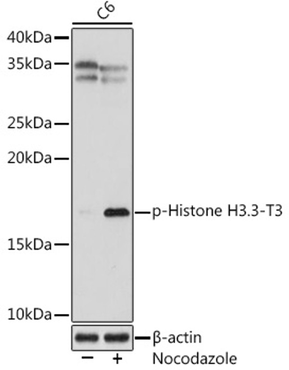
  • Western blot analysis of lysates from C6 cells, using Phospho-Histone H3. 3-T3 Rabbit monoclonal antibody (STJ11102645) at 1:1000 dilution. C6 cells were treated by Nocodazole (50 ng/mL) at 37 °C for 20 hours. Secondary antibody: HRP Goat Anti-Rabbit IgG (H+L) (STJS000856) at 1:10000 dilution. Lysates/proteins: 25 Mu g per lane. Blocking buffer: 3% BSA. Detection: ECL Enhanced Kit. Exposure time: 90s.
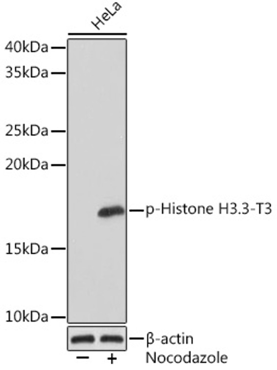
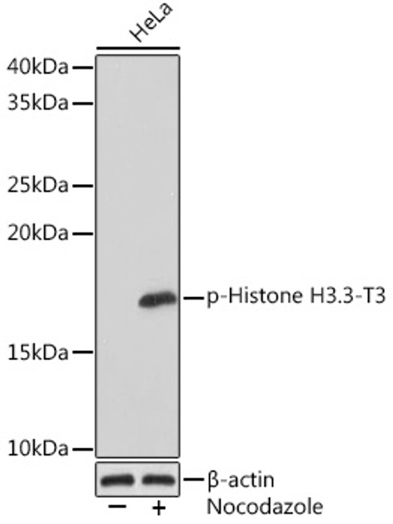
  • Western blot analysis of extracts of HeLa cells, using Phospho-Histone H3. 3-T3 rabbit monoclonal antibody (STJ11102645) at 1:1000 dilution. HeLa cells were treated by nocodazole (50 ng/mL) at 37 °C for 20 hours. Secondary antibody: HRP Goat Anti-rabbit IgG (H+L) at 1:10000 dilution. Lysates/proteins: 25ug per lane. Blocking buffer: 3% BSA. Detection: ECL Basic Kit. Exposure time: 1s.
  • Western blot analysis of lysates from HeLa cells, using Phospho-Histone H3. 3-T3 Rabbit monoclonal antibody (STJ11102645) at 1:1000 dilution. HeLa cells were treated by nocodazole (50 ng/mL) at 37 °C for 20 hours. Secondary antibody: HRP Goat Anti-Rabbit IgG (H+L) (STJS000856) at 1:10000 dilution. Lysates/proteins: 25 Mu g per lane. Blocking buffer: 3% BSA. Detection: ECL Basic Kit. Exposure time: 1s.
  • Western blot analysis of extracts of C6 cells, using Phospho-Histone H3. 3-T3 rabbit monoclonal antibody (STJ11102645) at 1:1000 dilution. C6 cells were treated by Nocodazole (50 ng/mL) at 37 °C for 20 hours. Secondary antibody: HRP Goat Anti-rabbit IgG (H+L) at 1:10000 dilution. Lysates/proteins: 25ug per lane. Blocking buffer: 3% BSA. Detection: ECL Enhanced Kit. Exposure time: 90s.
  • Western blot analysis of lysates from NIH/3T3 cells, using Phospho-Histone H3. 3-T3 Rabbit monoclonal antibody (STJ11102645) at 1:1000 dilution. NIH/3T3 cells were treated by Nocodazole (50 ng/mL) at 37 °C for 20 hours. Secondary antibody: HRP Goat Anti-Rabbit IgG (H+L) (STJS000856) at 1:10000 dilution. Lysates/proteins: 25 Mu g per lane. Blocking buffer: 3% BSA. Detection: ECL Enhanced Kit. Exposure time: 3min.

Anti-Phospho-Histone H3-T3 antibody [S5MR] (STJ11102645)

SKU:
STJ11102645

Shipping:
Free Shipping
Current Stock:
Host: Rabbit
Applications: WB
Reactivity: Human/Mouse/Rat
Note: STRICTLY FOR FURTHER SCIENTIFIC RESEARCH USE ONLY (RUO). MUST NOT TO BE USED IN DIAGNOSTIC OR THERAPEUTIC APPLICATIONS.
Short Description: Rabbit monoclonal antibody anti-Phospho-Histone H3-T3 is suitable for use in Western Blot research applications.
Clonality: Monoclonal
Clone ID: S5MR
Conjugation: Unconjugated
Isotype: IgG
Formulation: PBS with 0.02% Sodium Azide, 0.05% BSA, 50% Glycerol, pH7.3.
Purification: Affinity purification
Dilution Range: WB 1:500-1:1000
Storage Instruction: Store at-20°C for up to 1 year from the date of receipt, and avoid repeat freeze-thaw cycles.
Immunogen: A synthetic phosphorylated peptide around T3 of human Histone H3 (P84243).
Immunogen Sequence: MARTK
Background The protein encoded by this gene is a vesicle-bound, sodium-dependent phosphate transporter that is specifically expressed in the neuron-rich regions of the brain. It is preferentially associated with the membranes of synaptic vesicles and functions in glutamate transport. The protein shares 82% identity with the differentiation-associated Na-dependent inorganic phosphate cotransporter and they appear to form a distinct class within the Na+/Pi cotransporter family.

Information sourced from Uniprot.org

12 months for antibodies. 6 months for ELISA Kits. Please see website T&Cs for further guidance