| Host: |
Rabbit |
| Applications: |
WB/IF |
| Reactivity: |
Human/Mouse/Rat |
| Note: |
STRICTLY FOR FURTHER SCIENTIFIC RESEARCH USE ONLY (RUO). MUST NOT TO BE USED IN DIAGNOSTIC OR THERAPEUTIC APPLICATIONS. |
| Short Description: |
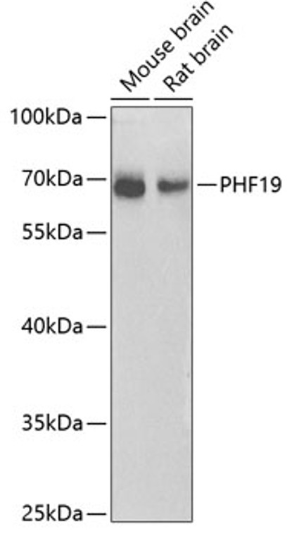
Rabbit polyclonal antibody anti-PHF19 (1-207) is suitable for use in Western Blot and Immunofluorescence research applications. |
| Clonality: |
Polyclonal |
| Conjugation: |
Unconjugated |
| Isotype: |
IgG |
| Formulation: |
PBS with 0.01% Thimerosal, 50% Glycerol, pH7.3. |
| Purification: |
Affinity purification |
| Dilution Range: |
WB 1:500-1:2000IF/ICC 1:50-1:100 |
| Storage Instruction: |
Store at-20°C for up to 1 year from the date of receipt, and avoid repeat freeze-thaw cycles. |
| Gene Symbol: |
PHF19 |
| Gene ID: |
26147 |
| Uniprot ID: |
PHF19_HUMAN |
| Immunogen Region: |
1-207 |
| Immunogen: |
Recombinant fusion protein containing a sequence corresponding to amino acids 1-207 of human PHF19 (NP_001009936.1). |
| Immunogen Sequence: |
MENRALDPGTRDSYGATSHL PNKGALAKVKNNFKDLMSKL TEGQYVLCRWTDGLYYLGKI KRVSSSKQSCLVTFEDNSKY WVLWKDIQHAGVPGEEPKCN ICLGKTSGPLNEILICGKCG LGYHQQCHIPIAGSADQPLL TPWFCRRCIFALAVRVSLPS SPVPASPASSSGADQRLPSQ SLSSKQKGHTWALETDSASA TVLGQDL |
| Tissue Specificity | Isoform 1 is expressed in thymus, heart, lung and kidney. Isoform 2 is predominantly expressed in placenta, skeletal muscle and kidney, whereas isoform 1 is predominantly expressed in liver and peripheral blood leukocytes. Overexpressed in many types of cancers, including colon, skin, lung, rectal, cervical, uterus, liver cancers, in cell lines derived from different stages of melanoma and in glioma cell lines. |
| Function | Polycomb group (PcG) protein that specifically binds histone H3 trimethylated at 'Lys-36' (H3K36me3) and recruits the PRC2 complex, thus enhancing PRC2 H3K27me3 methylation activity. Probably involved in the transition from an active state to a repressed state in embryonic stem cells: acts by binding to H3K36me3, a mark for transcriptional activation, and recruiting H3K36me3 histone demethylases RIOX1 or KDM2B, leading to demethylation of H3K36 and recruitment of the PRC2 complex that mediates H3K27me3 methylation, followed by de novo silencing. Recruits the PRC2 complex to CpG islands and contributes to embryonic stem cell self-renewal. Also binds histone H3 dimethylated at 'Lys-36' (H3K36me2). Isoform 1 and isoform 2 inhibit transcription from an HSV-tk promoter. |
| Protein Name | Phd Finger Protein 19Polycomb-Like Protein 3Hpcl3 |
| Database Links | Reactome: R-HSA-212300 |
| Cellular Localisation | NucleusLocalizes To Chromatin As Part Of The Prc2 Complex |
| Alternative Antibody Names | Anti-Phd Finger Protein 19 antibodyAnti-Polycomb-Like Protein 3 antibodyAnti-Hpcl3 antibodyAnti-PHF19 antibodyAnti-PCL3 antibody |
Information sourced from Uniprot.org
12 months for antibodies. 6 months for ELISA Kits. Please see website T&Cs for further guidance


