| Host: |
Rabbit |
| Applications: |
WB |
| Reactivity: |
Rat |
| Note: |
STRICTLY FOR FURTHER SCIENTIFIC RESEARCH USE ONLY (RUO). MUST NOT TO BE USED IN DIAGNOSTIC OR THERAPEUTIC APPLICATIONS. |
| Short Description: |
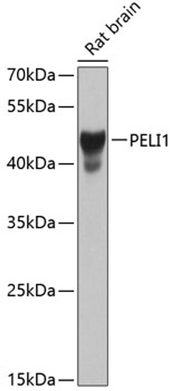
Rabbit polyclonal antibody anti-PELI1 (1-250) is suitable for use in Western Blot research applications. |
| Clonality: |
Polyclonal |
| Conjugation: |
Unconjugated |
| Isotype: |
IgG |
| Formulation: |
PBS with 0.02% Sodium Azide, 50% Glycerol, pH7.3. |
| Purification: |
Affinity purification |
| Dilution Range: |
WB 1:500-1:2000 |
| Storage Instruction: |
Store at-20°C for up to 1 year from the date of receipt, and avoid repeat freeze-thaw cycles. |
| Gene Symbol: |
PELI1 |
| Gene ID: |
57162 |
| Uniprot ID: |
PELI1_HUMAN |
| Immunogen Region: |
1-250 |
| Immunogen: |
Recombinant fusion protein containing a sequence corresponding to amino acids 1-250 of human PELI1 (NP_065702.2). |
| Immunogen Sequence: |
MFSPDQENHPSKAPVKYGEL IVLGYNGSLPNGDRGRRKSR FALFKRPKANGVKPSTVHIA CTPQAAKAISNKDQHSISYT LSRAQTVVVEYTHDSNTDMF QIGRSTESPIDFVVTDTVPG SQSNSDTQSVQSTISRFACR IICERNPPFTARIYAAGFDS SKNIFLGEKAAKWKTSDGQM DGLTTNGVLVMHPRNGFTED SKPGIWREISVCGNVFSLRE TRSAQQRGKMVEIETNQLQ |
| Tissue Specificity | Expressed at high levels in normal skin but decreased in keratinocytes from toxic epidermal necrolysis (TEN) patients (at protein level). |
| Post Translational Modifications | Phosphorylation by IRAK1 and IRAK4 enhances its E3 ligase activity. Sumoylated. |
| Function | E3 ubiquitin ligase catalyzing the covalent attachment of ubiquitin moieties onto substrate proteins. Involved in the TLR and IL-1 signaling pathways via interaction with the complex containing IRAK kinases and TRAF6. Mediates 'Lys-63'-linked polyubiquitination of IRAK1 allowing subsequent NF-kappa-B activation. Mediates 'Lys-48'-linked polyubiquitination of RIPK3 leading to its subsequent proteasome-dependent degradation.preferentially recognizes and mediates the degradation of the 'Thr-182' phosphorylated form of RIPK3. Negatively regulates necroptosis by reducing RIPK3 expression. Mediates 'Lys-63'-linked ubiquitination of RIPK1. |
| Protein Name | E3 Ubiquitin-Protein Ligase Pellino Homolog 1Pellino-1Pellino-Related Intracellular-Signaling MoleculeRing-Type E3 Ubiquitin Transferase Pellino Homolog 1 |
| Database Links | Reactome: R-HSA-5675482Reactome: R-HSA-9020702Reactome: R-HSA-937039Reactome: R-HSA-975144 |
| Alternative Antibody Names | Anti-E3 Ubiquitin-Protein Ligase Pellino Homolog 1 antibodyAnti-Pellino-1 antibodyAnti-Pellino-Related Intracellular-Signaling Molecule antibodyAnti-Ring-Type E3 Ubiquitin Transferase Pellino Homolog 1 antibodyAnti-PELI1 antibodyAnti-PRISM antibody |
Information sourced from Uniprot.org
12 months for antibodies. 6 months for ELISA Kits. Please see website T&Cs for further guidance


