| Host: |
Rabbit |
| Applications: |
WB/IF |
| Reactivity: |
Human/Rat |
| Note: |
STRICTLY FOR FURTHER SCIENTIFIC RESEARCH USE ONLY (RUO). MUST NOT TO BE USED IN DIAGNOSTIC OR THERAPEUTIC APPLICATIONS. |
| Short Description: |
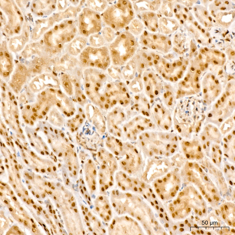
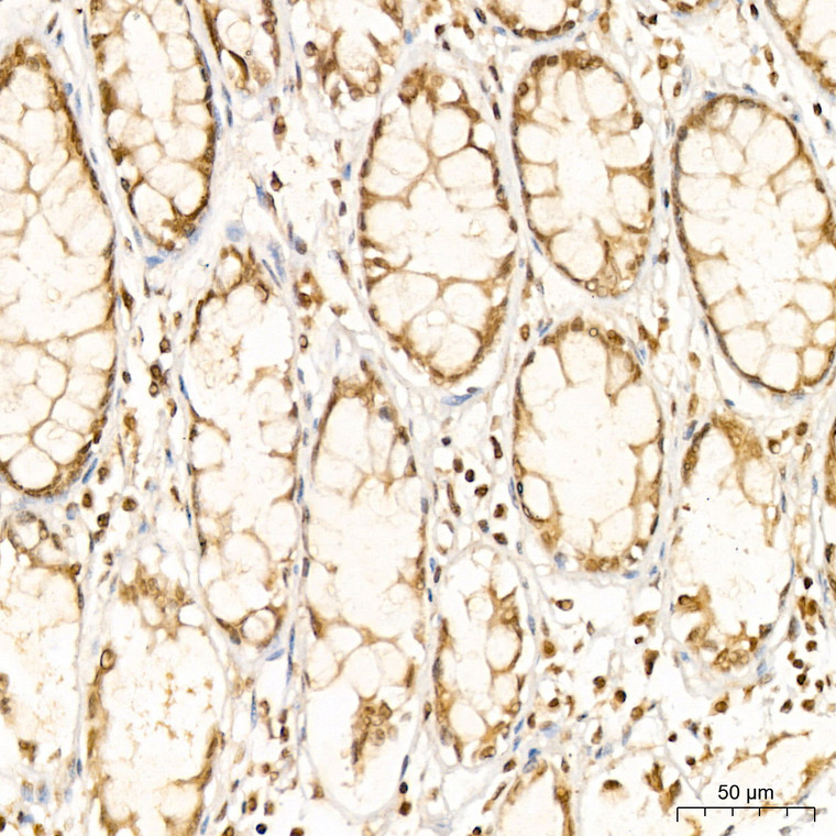
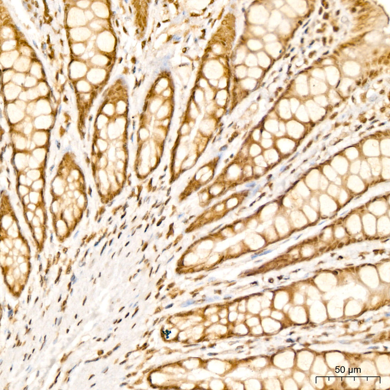
Rabbit polyclonal antibody anti-PARP3 (301-540) is suitable for use in Western Blot and Immunofluorescence research applications. |
| Clonality: |
Polyclonal |
| Conjugation: |
Unconjugated |
| Isotype: |
IgG |
| Formulation: |
PBS with 0.02% Sodium Azide, 50% Glycerol, pH7.3. |
| Purification: |
Affinity purification |
| Dilution Range: |
WB 1:500-1:1000IF/ICC 1:50-1:100 |
| Storage Instruction: |
Store at-20°C for up to 1 year from the date of receipt, and avoid repeat freeze-thaw cycles. |
| Gene Symbol: |
PARP3 |
| Gene ID: |
10039 |
| Uniprot ID: |
PARP3_HUMAN |
| Immunogen Region: |
301-540 |
| Immunogen: |
Recombinant fusion protein containing a sequence corresponding to amino acids 301-540 of human PARP3 (NP_001003931.3). |
| Immunogen Sequence: |
LAQALQAVSEQEKTVEEVPH PLDRDYQLLKCQLQLLDSGA PEYKVIQTYLEQTGSNHRCP TLQHIWKVNQEGEEDRFQAH SKLGNRKLLWHGTNMAVVAA ILTSGLRIMPHSGGRVGKGI YFASENSKSAGYVIGMKCGA HHVGYMFLGEVALGREHHIN TDNPSLKSPPPGFDSVIARG HTEPDPTQDTELELDGQQVV VPQGQPVPCPEFSSSTFSQS EYLIYQESQCRLRYLLEVH |
| Tissue Specificity | Widely expressed.the highest levels are in the kidney, skeletal muscle, liver, heart and spleen.also detected in pancreas, lung, placenta, brain, leukocytes, colon, small intestine, ovary, testis, prostate and thymus. |
| Post Translational Modifications | Auto-mono-ADP-ribosylated. |
| Function | Mono-ADP-ribosyltransferase that mediates mono-ADP-ribosylation of target proteins and plays a key role in the response to DNA damage. Mediates mono-ADP-ribosylation of glutamate, aspartate or lysine residues on target proteins. In contrast to PARP1 and PARP2, it is not able to mediate poly-ADP-ribosylation. Involved in DNA repair by mediating mono-ADP-ribosylation of a limited number of acceptor proteins involved in chromatin architecture and in DNA metabolism, such as histone H2B, XRCC5 and XRCC6. ADP-ribosylation follows DNA damage and appears as an obligatory step in a detection/signaling pathway leading to the reparation of DNA strand breaks. Involved in single-strand break repair by catalyzing mono-ADP-ribosylation of histone H2B on 'Glu-2' (H2BE2ADPr) of nucleosomes containing nicked DNA. Cooperates with the XRCC5-XRCC6 (Ku80-Ku70) heterodimer to limit end-resection thereby promoting accurate NHEJ. Suppresses G-quadruplex (G4) structures in response to DNA damage. Associates with a number of DNA repair factors and is involved in the response to exogenous and endogenous DNA strand breaks. Together with APLF, promotes the retention of the LIG4-XRCC4 complex on chromatin and accelerate DNA ligation during non-homologous end-joining (NHEJ). May link the DNA damage surveillance network to the mitotic fidelity checkpoint. Acts as a negative regulator of immunoglobulin class switch recombination, probably by controlling the level of AICDA /AID on the chromatin. In addition to proteins, also able to ADP-ribosylate DNA: mediates DNA mono-ADP-ribosylation of DNA strand break termini via covalent addition of a single ADP-ribose moiety to a 5'- or 3'-terminal phosphate residues in DNA containing multiple strand breaks. |
| Protein Name | Protein Mono-Adp-Ribosyltransferase Parp3Adp-Ribosyltransferase Diphtheria Toxin-Like 3Artd3Dna Adp-Ribosyltransferase Parp3Irt1Nad(+ Adp-Ribosyltransferase 3Adprt-3Poly Adp-Ribose Polymerase 3Parp-3Hparp-3PolyAdp-Ribose Synthase 3Padprt-3 |
| Cellular Localisation | NucleusChromosomeCytoplasmCytoskeletonMicrotubule Organizing CenterCentrosomeCentrioleAlmost Exclusively Localized In The Nucleus And Appears In Numerous Small Foci And A Small Number Of Larger Foci Whereas A Centrosomal Location Has Not Been DetectedIn Response To Dna DamageLocalizes To Sites Of Double-Strand BreakAlso Localizes To Single-Strand BreaksPreferentially Localized To The Daughter Centriole |
| Alternative Antibody Names | Anti-Protein Mono-Adp-Ribosyltransferase Parp3 antibodyAnti-Adp-Ribosyltransferase Diphtheria Toxin-Like 3 antibodyAnti-Artd3 antibodyAnti-Dna Adp-Ribosyltransferase Parp3 antibodyAnti-Irt1 antibodyAnti-Nad(+ Adp-Ribosyltransferase 3 antibodyAnti-Adprt-3 antibodyAnti-Poly Adp-Ribose Polymerase 3 antibodyAnti-Parp-3 antibodyAnti-Hparp-3 antibodyAnti-PolyAdp-Ribose Synthase 3 antibodyAnti-Padprt-3 antibodyAnti-PARP3 antibodyAnti-ADPRT3 antibodyAnti-ADPRTL3 antibody |
Information sourced from Uniprot.org
12 months for antibodies. 6 months for ELISA Kits. Please see website T&Cs for further guidance


