| Host: |
Rabbit |
| Applications: |
WB/IHC/IF |
| Reactivity: |
Human/Mouse/Rat |
| Note: |
STRICTLY FOR FURTHER SCIENTIFIC RESEARCH USE ONLY (RUO). MUST NOT TO BE USED IN DIAGNOSTIC OR THERAPEUTIC APPLICATIONS. |
| Short Description: |
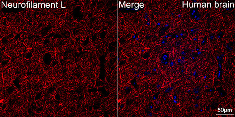
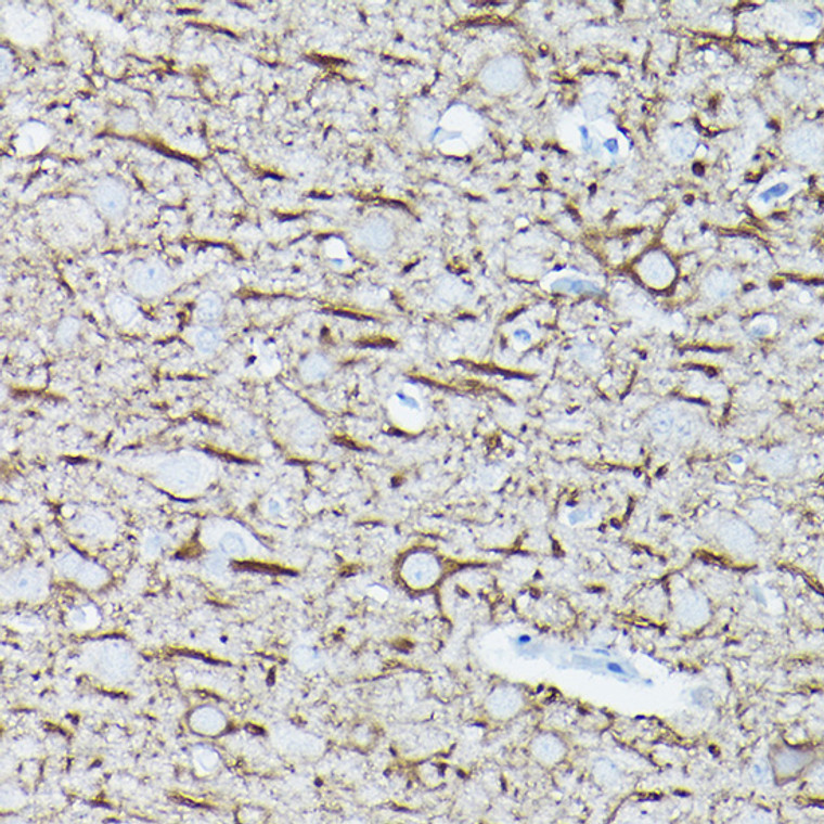
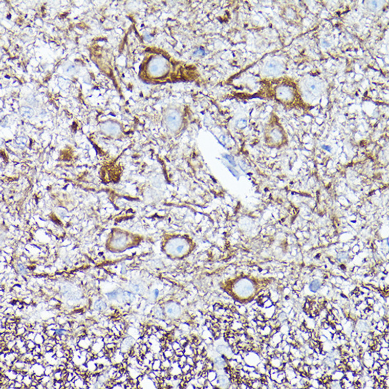
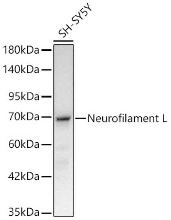
Rabbit monoclonal antibody anti-Neurofilament L (60-250) is suitable for use in Western Blot, Immunohistochemistry and Immunofluorescence research applications. |
| Clonality: |
Monoclonal |
| Clone ID: |
S0MR |
| Conjugation: |
Unconjugated |
| Isotype: |
IgG |
| Formulation: |
PBS with 0.01% Thimerosal, 50% Glycerol, pH7.3. |
| Purification: |
Affinity purification |
| Dilution Range: |
WB 1:500-1:1000IHC-P 1:100-1:500IF/ICC 1:50-1:200 |
| Storage Instruction: |
Store at-20°C for up to 1 year from the date of receipt, and avoid repeat freeze-thaw cycles. |
| Gene Symbol: |
NEFL |
| Gene ID: |
4747 |
| Uniprot ID: |
NFL_HUMAN |
| Immunogen Region: |
60-250 |
| Immunogen: |
Recombinant fusion protein containing a sequence corresponding to amino acids 60-250 of human Neurofilament L (NP_006149.2). |
| Immunogen Sequence: |
SSGSLMPSLENLDLSQVAAI SNDLKSIRTQEKAQLQDLND RFASFIERVHELEQQNKVLE AELLVLRQKHSEPSRFRALY EQEIRDLRLAAEDATNEKQA LQGEREGLEETLRNLQARYE EEVLSREDAEGRLMEARKGA DEAALARAELEKRIDSLMDE ISFLKKVHEEEIAELQAQIQ YAQISVEMDVT |
| Post Translational Modifications | O-glycosylated. Phosphorylated in the head and rod regions by the PKC kinase PKN1, leading to the inhibition of polymerization. Ubiquitinated in the presence of TRIM2 and UBE2D1. |
| Function | Neurofilaments usually contain three intermediate filament proteins: NEFL, NEFM, and NEFH which are involved in the maintenance of neuronal caliber. May additionally cooperate with the neuronal intermediate filament proteins PRPH and INA to form neuronal filamentous networks. |
| Protein Name | Neurofilament Light PolypeptideNf-L68 Kda Neurofilament ProteinNeurofilament Triplet L Protein |
| Database Links | Reactome: R-HSA-438066Reactome: R-HSA-442982Reactome: R-HSA-5673001Reactome: R-HSA-9609736Reactome: R-HSA-9617324Reactome: R-HSA-9620244 |
| Cellular Localisation | Cell ProjectionAxonCytoplasmCytoskeleton |
| Alternative Antibody Names | Anti-Neurofilament Light Polypeptide antibodyAnti-Nf-L antibodyAnti-68 Kda Neurofilament Protein antibodyAnti-Neurofilament Triplet L Protein antibodyAnti-NEFL antibodyAnti-NF68 antibodyAnti-NFL antibody |
Information sourced from Uniprot.org
12 months for antibodies. 6 months for ELISA Kits. Please see website T&Cs for further guidance


