| Host: |
Rabbit |
| Applications: |
WB/IHC/IF |
| Reactivity: |
Human/Mouse/Rat |
| Note: |
STRICTLY FOR FURTHER SCIENTIFIC RESEARCH USE ONLY (RUO). MUST NOT TO BE USED IN DIAGNOSTIC OR THERAPEUTIC APPLICATIONS. |
| Short Description: |
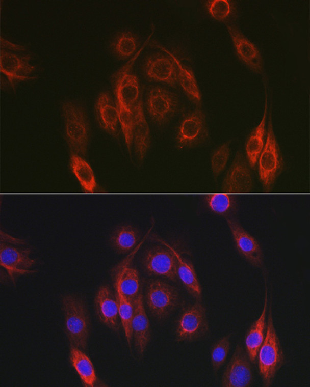
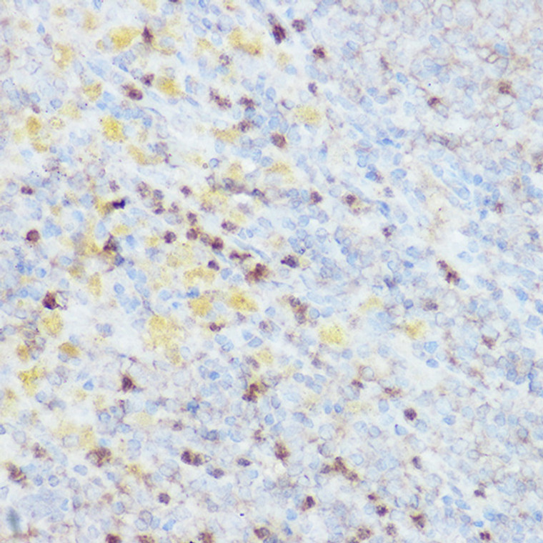
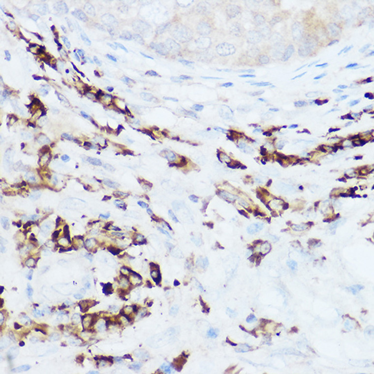
Rabbit polyclonal antibody anti-NEDD9 (390-560) is suitable for use in Western Blot, Immunohistochemistry and Immunofluorescence research applications. |
| Clonality: |
Polyclonal |
| Conjugation: |
Unconjugated |
| Isotype: |
IgG |
| Formulation: |
PBS with 0.01% Thimerosal, 50% Glycerol, pH7.3. |
| Purification: |
Affinity purification |
| Dilution Range: |
WB 1:500-1:1000IHC-P 1:50-1:200IF/ICC 1:50-1:200 |
| Storage Instruction: |
Store at-20°C for up to 1 year from the date of receipt, and avoid repeat freeze-thaw cycles. |
| Gene Symbol: |
NEDD9 |
| Gene ID: |
4739 |
| Uniprot ID: |
CASL_HUMAN |
| Immunogen Region: |
390-560 |
| Immunogen: |
Recombinant fusion protein containing a sequence corresponding to amino acids 390-560 of human NEDD9 (NP_006394.1). |
| Immunogen Sequence: |
SSLSASPAQDKRLFLDPDTA IERLQRLQQALEMGVSSLMA LVTTDWRCYGYMERHINEIR TAVDKVELFLKEYLHFVKGA VANAACLPELILHNKMKREL QRVEDSHQILSQTSHDLNEC SWSLNILAINKPQNKCDDLD RFVMVAKTVPDDAKQLTTTI NTNAEALFRPG |
| Tissue Specificity | Expressed in B-cells (at protein level). Expressed in the respiratory epithelium of the main bronchi to the bronchioles in the lungs (at protein level). High levels detected in kidney, lung, and placenta. Expressed in lymphocytes. |
| Post Translational Modifications | Cell cycle-regulated processing produces four isoforms: p115, p105, p65, and p55. Isoform p115 arises from p105 phosphorylation and appears later in the cell cycle. Isoform p55 arises from p105 as a result of cleavage at a caspase cleavage-related site and it appears specifically at mitosis. The p65 isoform is poorly detected. Polyubiquitinated by ITCH/AIP4, leading to proteasomal degradation. PTK2/FAK1 phosphorylates the protein at the YDYVHL motif (conserved among all cas proteins) following integrin stimulation. The SRC family kinases (FYN, SRC, LCK and CRK) are recruited to the phosphorylated sites and can phosphorylate other tyrosine residues. Ligation of either integrin beta-1 or B-cell antigen receptor on tonsillar B-cells and B-cell lines promotes tyrosine phosphorylation and both integrin and BCR-mediated tyrosine phosphorylation requires an intact actin network. Phosphorylation is required to recruit NEDD9 to T-cell receptor microclusters at the periphery of newly formed immunological synapses. In fibroblasts transformation with oncogene v-ABL results in an increase in tyrosine phosphorylation. Transiently phosphorylated following CD3 cross-linking and this phosphorylated form binds to CRKL and C3G. A mutant lacking the SH3 domain is phosphorylated upon CD3 cross-linking but not upon integrin beta-1 cross-linking. Tyrosine phosphorylation occurs upon stimulation of the G-protein coupled C1a calcitonin receptor. Calcitonin-stimulated tyrosine phosphorylation is mediated by calcium- and protein kinase C-dependent mechanisms and requires the integrity of the actin cytoskeleton. Phosphorylation at Ser-369 induces proteasomal degradation. Phosphorylated by LYN. Phosphorylation at Ser-780 by CSNK1D or CSNK1E, or phosphorylation of Thr-804 by CSNK1E enhances the interaction of NEDD9 with PLK1. |
| Function | Scaffolding protein which plays a central coordinating role for tyrosine-kinase-based signaling related to cell adhesion. As a focal adhesion protein, plays a role in embryonic fibroblast migration. May play an important role in integrin beta-1 or B cell antigen receptor (BCR) mediated signaling in B- and T-cells. Integrin beta-1 stimulation leads to recruitment of various proteins including CRKL and SHPTP2 to the tyrosine phosphorylated form. Promotes adhesion and migration of lymphocytes.as a result required for the correct migration of lymphocytes to the spleen and other secondary lymphoid organs. Plays a role in the organization of T-cell F-actin cortical cytoskeleton and the centralization of T-cell receptor microclusters at the immunological synapse. Negatively regulates cilia outgrowth in polarized cysts. Modulates cilia disassembly via activation of AURKA-mediated phosphorylation of HDAC6 and subsequent deacetylation of alpha-tubulin. Positively regulates RANKL-induced osteoclastogenesis. Required for the maintenance of hippocampal dendritic spines in the dentate gyrus and CA1 regions, thereby involved in spatial learning and memory. |
| Protein Name | Enhancer Of Filamentation 1Hef1Crk-Associated Substrate-Related ProteinCas-LCaslCas Scaffolding Protein Family Member 2Cass2Neural Precursor Cell Expressed Developmentally Down-Regulated Protein 9Nedd-9Renal Carcinoma Antigen Ny-Ren-12P105 Cleaved Into - Enhancer Of Filamentation 1 P55 |
| Cellular Localisation | CytoplasmCell CortexNucleusGolgi ApparatusCell ProjectionLamellipodiumCell JunctionFocal AdhesionCytoskeletonSpindle PoleCiliumCilium Basal BodyBasolateral Cell MembraneEnhancer Of Filamentation 1 P55: CytoplasmSpindle |
| Alternative Antibody Names | Anti-Enhancer Of Filamentation 1 antibodyAnti-Hef1 antibodyAnti-Crk-Associated Substrate-Related Protein antibodyAnti-Cas-L antibodyAnti-Casl antibodyAnti-Cas Scaffolding Protein Family Member 2 antibodyAnti-Cass2 antibodyAnti-Neural Precursor Cell Expressed Developmentally Down-Regulated Protein 9 antibodyAnti-Nedd-9 antibodyAnti-Renal Carcinoma Antigen Ny-Ren-12 antibodyAnti-P105 Cleaved Into - Enhancer Of Filamentation 1 P55 antibodyAnti-NEDD9 antibodyAnti-CASL antibody |
Information sourced from Uniprot.org
12 months for antibodies. 6 months for ELISA Kits. Please see website T&Cs for further guidance


