| Host: |
Rabbit |
| Applications: |
WB/IHC/IP |
| Reactivity: |
Human/Mouse/Rat |
| Note: |
STRICTLY FOR FURTHER SCIENTIFIC RESEARCH USE ONLY (RUO). MUST NOT TO BE USED IN DIAGNOSTIC OR THERAPEUTIC APPLICATIONS. |
| Short Description: |
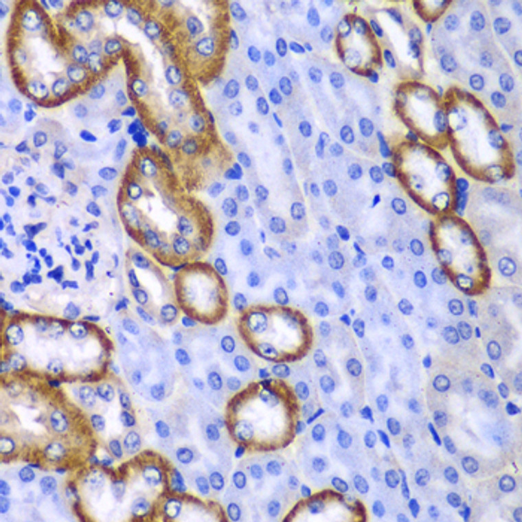
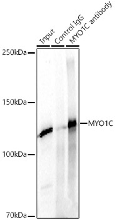
Rabbit polyclonal antibody anti-MYO1C (769-1028) is suitable for use in Western Blot, Immunohistochemistry and Immunoprecipitation research applications. |
| Clonality: |
Polyclonal |
| Conjugation: |
Unconjugated |
| Isotype: |
IgG |
| Formulation: |
PBS with 0.02% Sodium Azide, 50% Glycerol, pH7.3. |
| Purification: |
Affinity purification |
| Dilution Range: |
WB 1:500-1:2000IHC-P 1:50-1:200IP 1:50-1:200 |
| Storage Instruction: |
Store at-20°C for up to 1 year from the date of receipt, and avoid repeat freeze-thaw cycles. |
| Gene Symbol: |
MYO1C |
| Gene ID: |
4641 |
| Uniprot ID: |
MYO1C_HUMAN |
| Immunogen Region: |
769-1028 |
| Immunogen: |
Recombinant fusion protein containing a sequence corresponding to amino acids 769-1028 of human MYO1C (NP_203693.3). |
| Immunogen Sequence: |
ENAFFLDHVRTSFLLNLRRQ LPQNVLDTSWPTPPPALREA SELLRELCIKNMVWKYCRSI SPEWKQQLQQKAVASEIFKG KKDNYPQSVPRLFISTRLGT DEISPRVLQALGSEPIQYAV PVVKYDRKGYKPRSRQLLLT PNAVVIVEDAKVKQRIDYAN LTGISVSSLSDSLFVLHVQR ADNKQKGDVVLQSDHVIETL TKTALSANRVNSININQGSI TFAGGPGRDGTIDFTPGSE |
| Post Translational Modifications | Isoform 2 contains a N-acetylmethionine at position 1. |
| Function | Myosins are actin-based motor molecules with ATPase activity. Unconventional myosins serve in intracellular movements. Their highly divergent tails are presumed to bind to membranous compartments, which would be moved relative to actin filaments. Involved in glucose transporter recycling in response to insulin by regulating movement of intracellular GLUT4-containing vesicles to the plasma membrane. Component of the hair cell's (the sensory cells of the inner ear) adaptation-motor complex. Acts as a mediator of adaptation of mechanoelectrical transduction in stereocilia of vestibular hair cells. Binds phosphoinositides and links the actin cytoskeleton to cellular membranes. Isoform 3: Involved in regulation of transcription. Associated with transcriptional active ribosomal genes. Appears to cooperate with the WICH chromatin-remodeling complex to facilitate transcription. Necessary for the formation of the first phosphodiester bond during transcription initiation. |
| Protein Name | Unconventional Myosin-IcMyosin I BetaMmi-BetaMmib |
| Database Links | Reactome: R-HSA-1445148Reactome: R-HSA-2029482Reactome: R-HSA-5250924 O00159-3Reactome: R-HSA-9662360Reactome: R-HSA-9662361Reactome: R-HSA-9664422 |
| Cellular Localisation | CytoplasmNucleusCell CortexCell ProjectionStereocilium MembraneCytoplasmic VesicleRuffle MembraneColocalizes With Cabp1 And Cib1 At Cell MarginMembrane Ruffles And Punctate Regions On The Cell MembraneColocalizes In Adipocytes With Glut4 At Actin-Based MembranesColocalizes With Glut4 At Insulin-Induced Ruffles At The Cell MembraneLocalizes Transiently At Cell Membrane To Region Known To Be Enriched In Pip2Activation Of Phospholipase C Results In Its Redistribution To The CytoplasmColocalizes With Rna Polymerase IiTranslocates To Nuclear Speckles Upon Exposure To Inhibitors Of Rna Polymerase Ii TranscriptionIsoform 3: NucleusNucleoplasmNucleolusColocalizes With Rna Polymerase Ii In The NucleusColocalizes With Rna Polymerase I In NucleoliIn The NucleolusIs Localized Predominantly In Dense Fibrillar Component (Dfc) And In Granular Component (Gc)Accumulates Strongly In Dfc And Gc During Activation Of TranscriptionColocalizes With Transcription SitesColocalizes In The Granular Cortex At The Periphery Of The Nucleolus With Rps6Colocalizes In Nucleoplasm With Rps6 And Actin That Are In Contact With Rnp ParticlesColocalizes With Rps6 At The Nuclear Pore Level |
| Alternative Antibody Names | Anti-Unconventional Myosin-Ic antibodyAnti-Myosin I Beta antibodyAnti-Mmi-Beta antibodyAnti-Mmib antibodyAnti-MYO1C antibody |
Information sourced from Uniprot.org
12 months for antibodies. 6 months for ELISA Kits. Please see website T&Cs for further guidance


