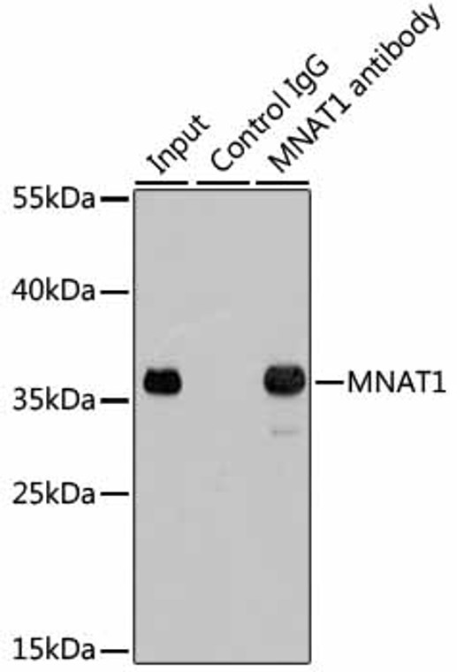
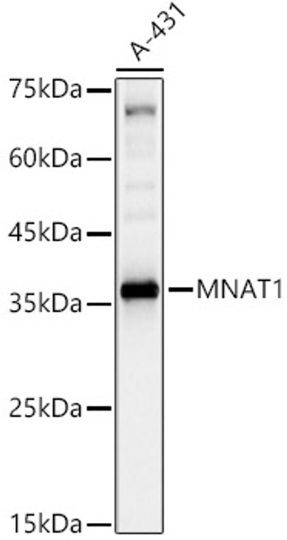
| Host: | Rabbit |
| Applications: | WB/IP |
| Reactivity: | Human/Mouse |
| Note: | STRICTLY FOR FURTHER SCIENTIFIC RESEARCH USE ONLY (RUO). MUST NOT TO BE USED IN DIAGNOSTIC OR THERAPEUTIC APPLICATIONS. |
| Short Description: | Rabbit polyclonal antibody anti-MNAT1 (1-309) is suitable for use in Western Blot and Immunoprecipitation research applications. |
| Clonality: | Polyclonal |
| Conjugation: | Unconjugated |
| Isotype: | IgG |
| Formulation: | PBS with 0.02% Sodium Azide, 50% Glycerol, pH7.3. |
| Purification: | Affinity purification |
| Dilution Range: | WB 1:500-1:1000IP 1:50-1:200 |
| Storage Instruction: | Store at-20°C for up to 1 year from the date of receipt, and avoid repeat freeze-thaw cycles. |
| Gene Symbol: | MNAT1 |
| Gene ID: | 4331 |
| Uniprot ID: | MAT1_HUMAN |
| Immunogen Region: | 1-309 |
| Immunogen: | Recombinant fusion protein containing a sequence corresponding to amino acids 1-309 of human MNAT1 (NP_002422.1). |
| Immunogen Sequence: | MDDQGCPRCKTTKYRNPSLK LMVNVCGHTLCESCVDLLFV RGAGNCPECGTPLRKSNFRV QLFEDPTVDKEVEIRKKVLK IYNKREEDFPSLREYNDFLE EVEEIVFNLTNNVDLDNTKK KMEIYQKENKDVIQKNKLKL TREQEELEEALEVERQENEQ RRLFIQKEEQLQQILKRKNK QAFLDELESSDLPVALLLAQ HKDRSTQLEMQLEKPKPVKP VTFSTGIKMGQHISLAPIH |
| Tissue Specificity | Highest levels in colon and testis. Moderate levels are present thymus, prostate, ovary, and small intestine. The lowest levels are found in spleen and leukocytes. |
| Function | Stabilizes the cyclin H-CDK7 complex to form a functional CDK-activating kinase (CAK) enzymatic complex. CAK activates the cyclin-associated kinases CDK1, CDK2, CDK4 and CDK6 by threonine phosphorylation. CAK complexed to the core-TFIIH basal transcription factor activates RNA polymerase II by serine phosphorylation of the repetitive C-terminal domain (CTD) of its large subunit (POLR2A), allowing its escape from the promoter and elongation of the transcripts. Involved in cell cycle control and in RNA transcription by RNA polymerase II. |
| Protein Name | Cdk-Activating Kinase Assembly Factor Mat1Cdk7/Cyclin-H Assembly FactorCyclin-G1-Interacting ProteinMenage A TroisRing Finger Protein 66Ring Finger Protein Mat1P35P36 |
| Database Links | Reactome: R-HSA-112382Reactome: R-HSA-113418Reactome: R-HSA-167152Reactome: R-HSA-167158Reactome: R-HSA-167160Reactome: R-HSA-167161Reactome: R-HSA-167162Reactome: R-HSA-167172Reactome: R-HSA-167200Reactome: R-HSA-167246Reactome: R-HSA-427413Reactome: R-HSA-5696395Reactome: R-HSA-674695Reactome: R-HSA-6781823Reactome: R-HSA-6781827Reactome: R-HSA-6782135Reactome: R-HSA-6782210Reactome: R-HSA-6796648Reactome: R-HSA-69202Reactome: R-HSA-69231Reactome: R-HSA-69273Reactome: R-HSA-69656Reactome: R-HSA-72086Reactome: R-HSA-73762Reactome: R-HSA-73772Reactome: R-HSA-73776Reactome: R-HSA-73779Reactome: R-HSA-73863Reactome: R-HSA-75953Reactome: R-HSA-75955Reactome: R-HSA-76042Reactome: R-HSA-77075Reactome: R-HSA-8939236 |
| Cellular Localisation | Nucleus |
| Alternative Antibody Names | Anti-Cdk-Activating Kinase Assembly Factor Mat1 antibodyAnti-Cdk7/Cyclin-H Assembly Factor antibodyAnti-Cyclin-G1-Interacting Protein antibodyAnti-Menage A Trois antibodyAnti-Ring Finger Protein 66 antibodyAnti-Ring Finger Protein Mat1 antibodyAnti-P35 antibodyAnti-P36 antibodyAnti-MNAT1 antibodyAnti-CAP35 antibodyAnti-MAT1 antibodyAnti-RNF66 antibody |
Information sourced from Uniprot.org
12 months for antibodies. 6 months for ELISA Kits. Please see website T&Cs for further guidance


