| Host: |
Rabbit |
| Applications: |
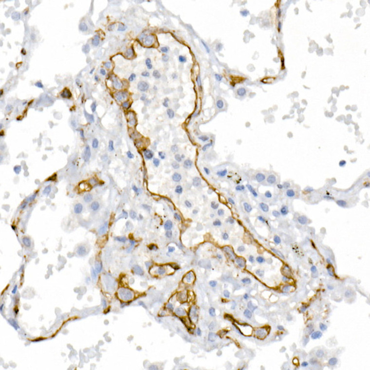
WB/IHC/IF |
| Reactivity: |
Human/Mouse/Rat |
| Note: |
STRICTLY FOR FURTHER SCIENTIFIC RESEARCH USE ONLY (RUO). MUST NOT TO BE USED IN DIAGNOSTIC OR THERAPEUTIC APPLICATIONS. |
| Short Description: |
Rabbit polyclonal antibody anti-MCAM (26-646) is suitable for use in Western Blot, Immunohistochemistry and Immunofluorescence research applications. |
| Clonality: |
Polyclonal |
| Conjugation: |
Unconjugated |
| Isotype: |
IgG |
| Formulation: |
PBS with 0.05% Proclin300, 50% Glycerol, pH7.3. |
| Purification: |
Affinity purification |
| Dilution Range: |
WB 1:100-1:500IHC-P 1:50-1:200IF/ICC 1:50-1:200 |
| Storage Instruction: |
Store at-20°C for up to 1 year from the date of receipt, and avoid repeat freeze-thaw cycles. |
| Gene Symbol: |
MCAM |
| Gene ID: |
4162 |
| Uniprot ID: |
MUC18_HUMAN |
| Immunogen Region: |
26-646 |
| Immunogen: |
Recombinant fusion protein containing a sequence corresponding to amino acids 26-646 of human CD146/MCAM (NP_006491.2). |
| Immunogen Sequence: |
GEAEQPAPELVEVEVGSTAL LKCGLSQSQGNLSHVDWFSV HKEKRTLIFRVRQGQGQSEP GEYEQRLSLQDRGATLALTQ VTPQDERIFLCQGKRPRSQE YRIQLRVYKAPEEPNIQVNP LGIPVNSKEPEEVATCVGRN GYPIPQVIWYKNGRPLKEEK NRVHIQSSQTVESSGLYTLQ SILKAQLVKEDKDAQFYCEL NYRLPSGNHMKESREVTVPV FYPTEKVWLEVEPVGMLKE |
| Tissue Specificity | Detected in endothelial cells in vascular tissue throughout the body. May appear at the surface of neural crest cells during their embryonic migration. Appears to be limited to vascular smooth muscle in normal adult tissues. Associated with tumor progression and the development of metastasis in human malignant melanoma. Expressed most strongly on metastatic lesions and advanced primary tumors and is only rarely detected in benign melanocytic nevi and thin primary melanomas with a low probability of metastasis. |
| Function | Plays a role in cell adhesion, and in cohesion of the endothelial monolayer at intercellular junctions in vascular tissue. Its expression may allow melanoma cells to interact with cellular elements of the vascular system, thereby enhancing hematogeneous tumor spread. Could be an adhesion molecule active in neural crest cells during embryonic development. Acts as surface receptor that triggers tyrosine phosphorylation of FYN and PTK2/FAK1, and a transient increase in the intracellular calcium concentration. |
| Protein Name | Cell Surface Glycoprotein Muc18Cell Surface Glycoprotein P1h12Melanoma Cell Adhesion MoleculeMelanoma-Associated Antigen A32Melanoma-Associated Antigen Muc18S-Endo 1 Endothelial-Associated AntigenCd Antigen Cd146 |
| Database Links | Reactome: R-HSA-8980692Reactome: R-HSA-9013026Reactome: R-HSA-9013106Reactome: R-HSA-9013149Reactome: R-HSA-9013404Reactome: R-HSA-9013405Reactome: R-HSA-9013408Reactome: R-HSA-9013423Reactome: R-HSA-9035034 |
| Cellular Localisation | MembraneSingle-Pass Type I Membrane Protein |
| Alternative Antibody Names | Anti-Cell Surface Glycoprotein Muc18 antibodyAnti-Cell Surface Glycoprotein P1h12 antibodyAnti-Melanoma Cell Adhesion Molecule antibodyAnti-Melanoma-Associated Antigen A32 antibodyAnti-Melanoma-Associated Antigen Muc18 antibodyAnti-S-Endo 1 Endothelial-Associated Antigen antibodyAnti-Cd Antigen Cd146 antibodyAnti-MCAM antibodyAnti-MUC18 antibody |
Information sourced from Uniprot.org
12 months for antibodies. 6 months for ELISA Kits. Please see website T&Cs for further guidance


![Western blot analysis of lysates from wild type (WT) and CD146/CD146/MCAM knockout (KO) HeLa cells, using [KO Validated] CD146/MCAM Rabbit polyclonal antibody (STJ11100869) at 1:1000 dilution. Secondary antibody: HRP Goat Anti-Rabbit IgG (H+L) (STJS000856) at 1:10000 dilution. Lysates/proteins: 25 Mu g per lane. Blocking buffer: 3% nonfat dry milk in TBST. Detection: ECL Basic Kit. Exposure time: 1s. Western blot analysis of lysates from wild type (WT) and CD146/CD146/MCAM knockout (KO) HeLa cells, using [KO Validated] CD146/MCAM Rabbit polyclonal antibody (STJ11100869) at 1:1000 dilution. Secondary antibody: HRP Goat Anti-Rabbit IgG (H+L) (STJS000856) at 1:10000 dilution. Lysates/proteins: 25 Mu g per lane. Blocking buffer: 3% nonfat dry milk in TBST. Detection: ECL Basic Kit. Exposure time: 1s.](https://cdn11.bigcommerce.com/s-zso2xnchw9/images/stencil/300x300/products/89803/358270/STJ11100869_1__94433.1713122612.jpg?c=1)

