| Host: |
Rabbit |
| Applications: |
WB/IHC/IF |
| Reactivity: |
Human/Mouse/Rat |
| Note: |
STRICTLY FOR FURTHER SCIENTIFIC RESEARCH USE ONLY (RUO). MUST NOT TO BE USED IN DIAGNOSTIC OR THERAPEUTIC APPLICATIONS. |
| Short Description: |
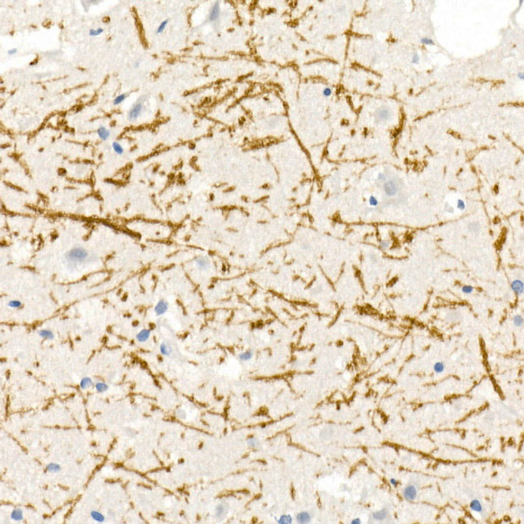
Rabbit polyclonal antibody anti-Myelin Basic protein (80-165) is suitable for use in Western Blot, Immunohistochemistry and Immunofluorescence research applications. |
| Clonality: |
Polyclonal |
| Conjugation: |
Unconjugated |
| Isotype: |
IgG |
| Formulation: |
PBS with 0.01% Thimerosal, 50% Glycerol, pH7.3. |
| Purification: |
Affinity purification |
| Dilution Range: |
WB 1:500-1:1000IHC-P 1:50-1:200IF/ICC 1:50-1:200 |
| Storage Instruction: |
Store at-20°C for up to 1 year from the date of receipt, and avoid repeat freeze-thaw cycles. |
| Gene Symbol: |
MBP |
| Gene ID: |
4155 |
| Uniprot ID: |
MBP_HUMAN |
| Immunogen Region: |
80-165 |
| Immunogen: |
Recombinant fusion protein containing a sequence corresponding to amino acids 80-165 of human Myelin Basic Protein (NP_001020272.1). |
| Immunogen Sequence: |
AHPADPGSRPHLIRLFSRDA PGREDNTFKDRPSESDELQT IQEDSAATSESLDVMASQKR PSQRHGSKYLATASTMDHAR HGFLPR |
| Tissue Specificity | MBP isoforms are found in both the central and the peripheral nervous system, whereas Golli-MBP isoforms are expressed in fetal thymus, spleen and spinal cord, as well as in cell lines derived from the immune system. |
| Post Translational Modifications | Several charge isomers of MBP.C1 (the most cationic, least modified, and most abundant form), C2, C3, C4, C5, C6, C7, C8-A and C8-B (the least cationic form).are produced as a result of optional PTM, such as phosphorylation, deamidation of glutamine or asparagine, arginine citrullination and methylation. C8-A and C8-B contain each two mass isoforms termed C8-A(H), C8-A(L), C8-B(H) and C8-B(L), (H) standing for higher and (L) for lower molecular weight. C3, C4 and C5 are phosphorylated. The ratio of methylated arginine residues decreases during aging, making the protein more cationic. The N-terminal alanine is acetylated (isoform 3, isoform 4, isoform 5 and isoform 6). Arg-241 was found to be 6% monomethylated and 60% symmetrically dimethylated. Proteolytically cleaved in B cell lysosomes by cathepsin CTSG which degrades the major immunogenic MBP epitope and prevents the activation of MBP-specific autoreactive T cells. Phosphorylated by TAOK2, VRK2, MAPK11, MAPK12, MAPK14 and MINK1. |
| Function | The classic group of MBP isoforms (isoform 4-isoform 14) are with PLP the most abundant protein components of the myelin membrane in the CNS. They have a role in both its formation and stabilization. The smaller isoforms might have an important role in remyelination of denuded axons in multiple sclerosis. The non-classic group of MBP isoforms (isoform 1-isoform 3/Golli-MBPs) may preferentially have a role in the early developing brain long before myelination, maybe as components of transcriptional complexes, and may also be involved in signaling pathways in T-cells and neural cells. Differential splicing events combined with optional post-translational modifications give a wide spectrum of isomers, with each of them potentially having a specialized function. Induces T-cell proliferation. |
| Protein Name | Myelin Basic ProteinMbpMyelin A1 ProteinMyelin Membrane Encephalitogenic Protein |
| Database Links | Reactome: R-HSA-9619665 |
| Cellular Localisation | Myelin MembranePeripheral Membrane ProteinCytoplasmic SideCytoplasmic Side Of MyelinIsoform 3: NucleusTargeted To Nucleus In Oligodendrocytes |
| Alternative Antibody Names | Anti-Myelin Basic Protein antibodyAnti-Mbp antibodyAnti-Myelin A1 Protein antibodyAnti-Myelin Membrane Encephalitogenic Protein antibodyAnti-MBP antibody |
Information sourced from Uniprot.org
12 months for antibodies. 6 months for ELISA Kits. Please see website T&Cs for further guidance


