| Host: |
Rabbit |
| Applications: |
WB/IHC/IF/ELISA |
| Reactivity: |
Human/Mouse/Rat |
| Note: |
STRICTLY FOR FURTHER SCIENTIFIC RESEARCH USE ONLY (RUO). MUST NOT TO BE USED IN DIAGNOSTIC OR THERAPEUTIC APPLICATIONS. |
| Short Description: |
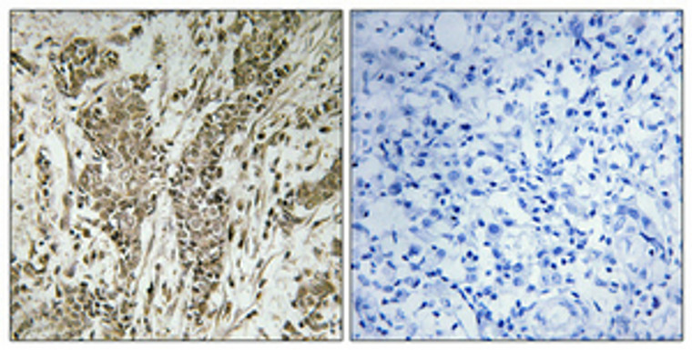
Rabbit polyclonal antibody anti-Membrane-associated guanylate kinase, WW and PDZ domain-containing protein 2 (221-270 aa) is suitable for use in Western Blot, Immunohistochemistry, Immunofluorescence and ELISA research applications. |
| Clonality: |
Polyclonal |
| Conjugation: |
Unconjugated |
| Isotype: |
IgG |
| Formulation: |
Liquid in PBS containing 50% Glycerol, 0.5% BSA and 0.02% Sodium Azide. |
| Purification: |
The antibody was affinity-purified from rabbit antiserum by affinity-chromatography using epitope-specific immunogen. |
| Concentration: |
1 mg/mL |
| Dilution Range: |
WB 1:500-1:2000IHC 1:100-1:300ELISA 1:20000IF 1:50-200 |
| Storage Instruction: |
Store at-20°C for up to 1 year from the date of receipt, and avoid repeat freeze-thaw cycles. |
| Gene Symbol: |
MAGI2 |
| Gene ID: |
9863 |
| Uniprot ID: |
MAGI2_HUMAN |
| Immunogen Region: |
221-270 aa |
| Specificity: |
MAGI-2 Polyclonal Antibody detects endogenous levels of MAGI-2 protein. |
| Immunogen: |
The antiserum was produced against synthesized peptide derived from the human MAGI2 at the amino acid range 221-270 |
| Function | Seems to act as a scaffold molecule at synaptic junctions by assembling neurotransmitter receptors and cell adhesion proteins. Plays a role in nerve growth factor (NGF)-induced recruitment of RAPGEF2 to late endosomes and neurite outgrowth. May play a role in regulating activin-mediated signaling in neuronal cells. Enhances the ability of PTEN to suppress AKT1 activation. Plays a role in receptor-mediated clathrin-dependent endocytosis which is required for ciliogenesis. |
| Protein Name | Membrane-Associated Guanylate Kinase - Ww And Pdz Domain-Containing Protein 2Atrophin-1-Interacting Protein 1Aip-1Atrophin-1-Interacting Protein AMembrane-Associated Guanylate Kinase Inverted 2Magi-2 |
| Database Links | Reactome: R-HSA-373753 |
| Cellular Localisation | CytoplasmLate EndosomeSynapseSynaptosomeCell MembranePeripheral Membrane ProteinCytoskeletonMicrotubule Organizing CenterCentrosomeCell ProjectionCiliumCentriolePhotoreceptor Inner SegmentPhotoreceptor Outer SegmentLocalized Diffusely In The Cytoplasm Before Nerve Growth Factor (Ngf) StimulationRecruited To Late Endosomes After Ngf StimulationMembrane-Associated In Synaptosomes |
| Alternative Antibody Names | Anti-Membrane-Associated Guanylate Kinase - Ww And Pdz Domain-Containing Protein 2 antibodyAnti-Atrophin-1-Interacting Protein 1 antibodyAnti-Aip-1 antibodyAnti-Atrophin-1-Interacting Protein A antibodyAnti-Membrane-Associated Guanylate Kinase Inverted 2 antibodyAnti-Magi-2 antibodyAnti-MAGI2 antibodyAnti-ACVRINP1 antibodyAnti-AIP1 antibodyAnti-KIAA0705 antibody |
Information sourced from Uniprot.org
12 months for antibodies. 6 months for ELISA Kits. Please see website T&Cs for further guidance


